


Semax Benefits
What Research Shows
Introduction
Semax is a synthetic regulatory peptide studied for its potential effects on cognitive performance, neurotrophin signaling, brain-network activity, and neurological responses to ischemic or cellular stress.¹–⁶
Unlike peptides that act through one clearly established receptor pathway, Semax appears to influence several interconnected neurological systems. Its proposed research benefits are therefore linked to a broader profile involving BDNF and other neurotrophins, monoamine signaling, gene expression, and cellular stress responses.
This article reviews the major research benefits associated with Semax, including attention and memory, neural plasticity, brain-network connectivity, cerebral-ischemia research, neuroimmune signaling, and neuronal resilience.
As discussed in How Does Semax Work?, the complete mechanism has not been established. Most evidence remains preclinical, while the available human studies are relatively small and have limited independent replication.
Summary Table: Semax Benefits and Evidence
| Benefit/ Research Area | Evidence Level | Study Type | Notes |
| 1. Attention and memory research | Limited human; moderate preclinical | Small human studies and animal behavioral models | Older studies reported changes in attention, short-term memory, and learning |
| 2. Neurotrophin and neural-plasticity signaling | Strong mechanistic preclinical | Rodent and cell studies | Semax altered BDNF, TrkB, NGF, and related signaling markers |
| 3. Brain-network connectivity | Preliminary human | Resting-state fMRI studies | Small studies reported changes in default-mode and regional functional connectivity |
| 4. Cerebral-ischemia and neurological-recovery research | Limited human; substantial preclinical | Human observational studies and animal ischemia models | Studied in acute stroke, rehabilitation, infarct models, and post-ischemic learning |
| 5. Neuroimmune and vascular-response research | Mechanistic preclinical | Transcriptomic and protein-expression studies | Altered genes related to inflammation, neurotransmission, and vascular function |
| 6. Neuronal resilience and cellular-stress research | Moderate preclinical | Cultured-neuron and animal studies | Associated with calcium regulation, mitochondrial stability, and oxidative-stress pathways |
1. Attention and Memory Research
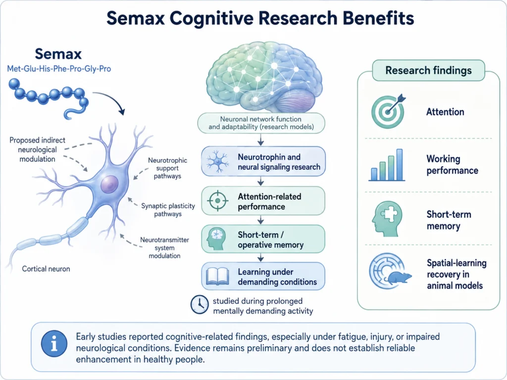
Semax is often described as a nootropic research peptide because several early studies examined its effects on attention, working performance, and memory.
A small 1996 study evaluated Semax in healthy volunteers completing prolonged periods of mentally demanding activity. The researchers reported improvements in measures of attention and short-term memory, with some effects becoming more apparent as participants became fatigued.¹
A broader 1997 report also described improvements in operative memory and attention under demanding conditions.² However, these early studies were small, and their reporting and methodology do not provide the same level of evidence as a modern, large, independently replicated clinical trial.
Animal studies provide additional support for cognitive research. In rats with ischemic lesions to the prefrontal cortex, Semax administration was associated with recovery of learning performance in a spatial-memory task.³ Other rodent studies have reported changes in passive-avoidance learning and memory-related behavior.
These findings suggest that Semax may be particularly relevant to cognition under conditions of fatigue, injury, hypoxia, or impaired neurological function. They do not establish that Semax reliably enhances memory or attention in healthy people.
Why this matters:
Semax has both human and animal evidence related to attention and memory, but the direct human evidence is limited. It is more accurate to describe Semax as a peptide studied in cognitive-performance models than as a proven cognitive enhancer.
2. Neurotrophin and Neural-Plasticity Signaling
One of the strongest areas of Semax research involves neurotrophins.
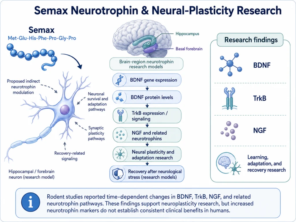
Neurotrophins are signaling proteins involved in neuronal survival, adaptation, development, and synaptic plasticity. The most frequently studied Semax-related neurotrophin is brain-derived neurotrophic factor, or BDNF.
Rodent studies have reported that Semax alters:
- BDNF gene expression
- BDNF protein levels
- Expression or activation of the BDNF receptor TrkB
- Nerve growth factor, or NGF
- Other neurotrophins and their receptors
In the rat hippocampus, Semax produced time-dependent changes in BDNF and TrkB expression and signaling.⁴ Another study reported increased BDNF protein in the rat basal forebrain.⁵
These molecular effects provide a possible biological connection between Semax and research on learning, neural adaptation, and recovery following neurological stress.
However, an increase in a neurotrophin marker is not itself a clinical benefit. BDNF levels vary by brain region, timing, and experimental condition, and it has not been established that Semax produces consistent, meaningful neuroplasticity outcomes in humans.
Why this matters:
Neurotrophin modulation is one of the most biologically supported reasons for studying Semax. It may help researchers investigate how BDNF-related pathways respond to learning, injury, or reduced blood flow, but direct human benefits remain uncertain.
3. Brain-Network Connectivity Research
Human imaging studies have examined whether Semax produces measurable changes in communication between brain regions.
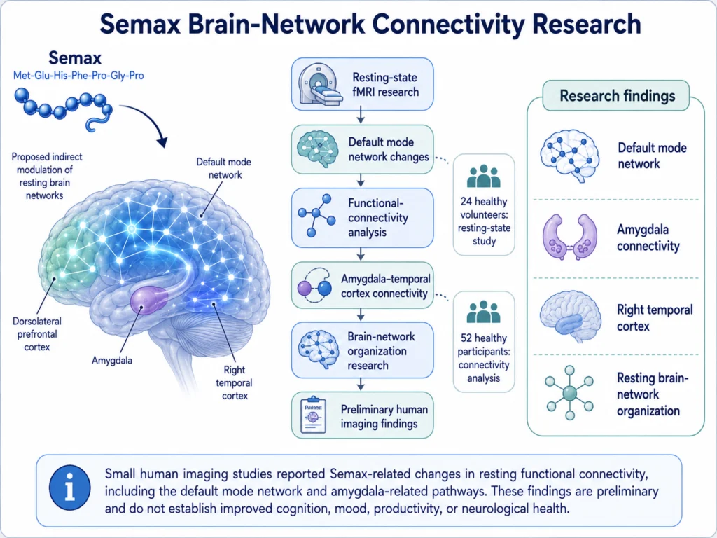
A small resting-state functional MRI study included 24 healthy volunteers who received Semax or placebo. Researchers reported changes in the default mode network following Semax administration.⁶
The default mode network is a group of connected brain regions associated with internally directed thought, memory-related processing, and integration of information when a person is not focused on a specific external task.
A later functional-connectivity analysis involving 52 healthy participants examined connections involving the amygdala and dorsolateral prefrontal cortex. The study reported Semax-related changes in connectivity between the right amygdala and regions of the right temporal cortex.⁷
These studies demonstrate that Semax can be investigated through measurable changes in resting brain-network organization. They do not establish that the reported connectivity changes improve cognition, mood, productivity, or neurological health.
Why this matters:
Brain-imaging research provides preliminary human evidence that Semax may influence functional communication between neurological regions. However, changes on an fMRI scan should not automatically be interpreted as a beneficial cognitive outcome.
4. Cerebral-Ischemia and Neurological-Recovery Research
A large portion of the Semax literature involves cerebral ischemia, which occurs when blood flow to part of the brain is reduced or interrupted.
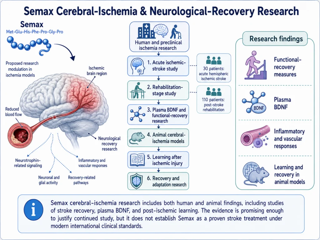
Early human studies examined Semax alongside conventional care in patients with ischemic stroke. A 1997 study included 30 patients receiving Semax during the acute phase of hemispheric ischemic stroke and compared their outcomes with a larger conventionally treated group.⁸
A later study evaluated 110 patients during rehabilitation following ischemic stroke. Semax administration was associated with increased plasma BDNF and reported improvements in motor and functional-recovery measures, particularly when combined with early rehabilitation.⁹
These studies are relevant because they examined functional outcomes in people rather than molecular markers alone. However, they were conducted within a limited clinical-research setting and have not been broadly reproduced in large international trials.
The preclinical literature is more extensive. In animal ischemia models, Semax has been associated with:
- Reduced infarct-related tissue changes
- Improved learning after ischemic injury
- Changes in neuronal and glial-cell activity
- Increased expression of neurotrophins
- Modification of inflammatory and vascular responses
- Changes in proteins related to cell death and neurological recovery
For example, Semax restored spatial-learning performance in rats after ischemic injury to the prefrontal cortex.³ Other studies found changes in brain-cell morphology, proliferation, gene expression, and protein markers following cerebral ischemia.
Why this matters:
Cerebral ischemia is one of the most developed areas of Semax research and includes both human and preclinical findings. The evidence is promising enough to justify continued study, but it does not establish Semax as a proven stroke treatment under modern international clinical standards.
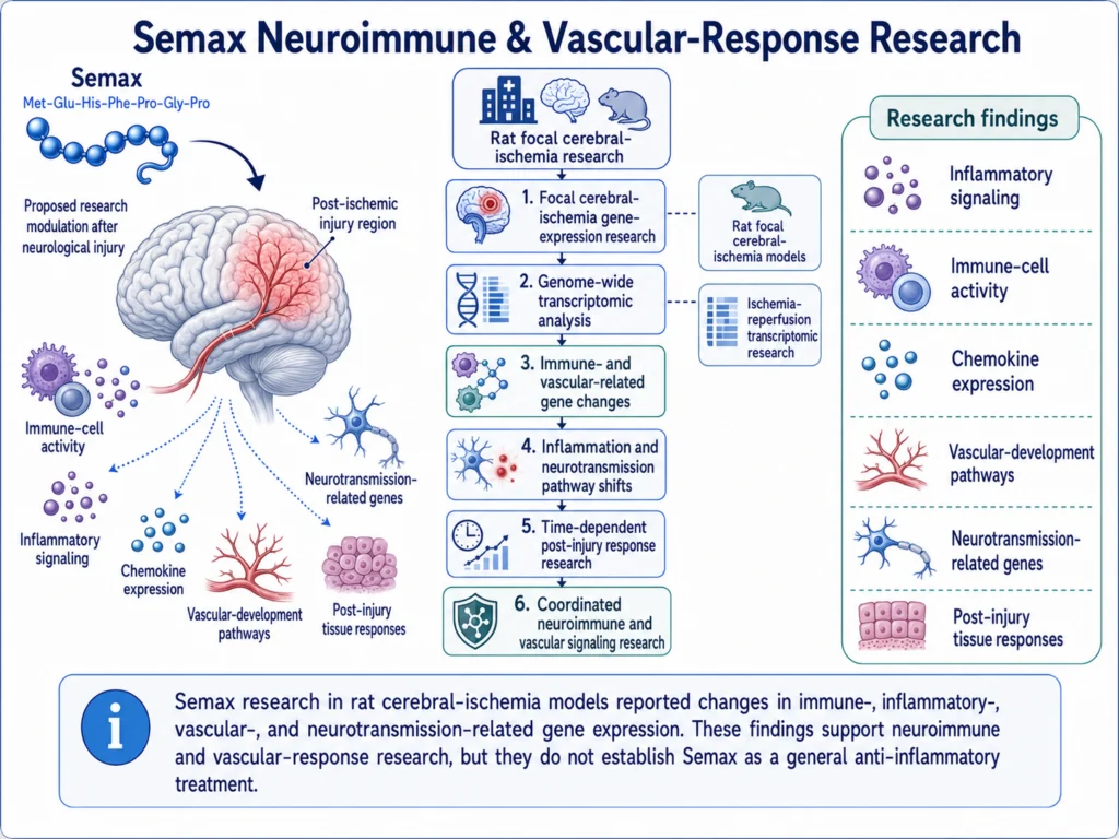
5. Neuroimmune and Vascular-Response Research
Neurological injury affects more than neurons. It also activates immune cells, inflammatory mediators, vascular pathways, and tissue-repair processes.
Genome-wide research in rats found that Semax altered the expression of genes related to immune activity and vascular function following focal cerebral ischemia.¹⁰ The effects changed over time, suggesting that Semax may influence different stages of the response to neurological injury.
A later transcriptomic study found that ischemia-reperfusion increased the expression of inflammation-related genes and suppressed genes involved in neurotransmission. Semax partially shifted these patterns in the opposite direction.¹¹
This research suggests potential effects involving:
- Inflammatory signaling
- Immune-cell activity
- Chemokine expression
- Vascular-development pathways
- Neurotransmission-related genes
- Post-injury tissue responses
The findings do not mean that Semax acts as a general anti-inflammatory agent. They were observed in specific rat models of cerebral ischemia and may reflect coordinated changes across several biological systems.
Why this matters:
Semax’s research profile extends beyond direct neuronal signaling. Its effects on immune- and vascular-related gene expression may help researchers study how the brain coordinates inflammation, blood-vessel responses, and neurological recovery after injury.
6. Neuronal Resilience and Cellular-Stress Research
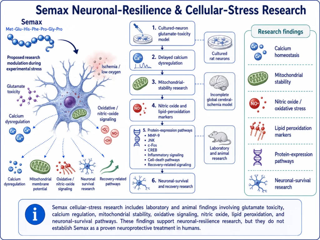
Semax has also been studied for its potential to help neurons maintain function during experimental stress.
Excessive glutamate activity can disturb intracellular calcium regulation, damage mitochondrial function, and contribute to neuronal death. In cultured rat neurons exposed to glutamate toxicity, Semax delayed calcium dysregulation and the loss of mitochondrial membrane potential. It was also associated with increased neuronal survival in that laboratory model.¹²
Animal research has examined additional stress-related pathways. In rats with incomplete global cerebral ischemia, Semax reduced the ischemia-associated increase in nitric oxide production and altered markers related to lipid peroxidation.¹³
Protein-expression studies have also reported Semax-associated changes involving:
- MMP-9
- JNK
- c-Fos
- CREB
- Inflammatory signaling
- Cell-death pathways
- Recovery-related signaling
Together, these findings suggest that Semax may influence how neurons and surrounding cells respond to excitotoxicity, reduced oxygen, oxidative stress, and mitochondrial instability.
These are laboratory and animal findings. They do not establish that Semax protects the human brain from injury or prevents neurological disease.
Why this matters:
Cellular-stress models help connect Semax’s molecular effects with measurable neuronal-survival outcomes. They also show why Semax is studied as a multi-pathway peptide rather than as a compound affecting only BDNF or cognition.
Semax’s Multi-Pathway Neurological Profile
Unlike peptides that produce one primary endocrine effect, Semax is studied across several overlapping neurological systems:
- Attention, learning, and memory-related performance
- BDNF, TrkB, and broader neurotrophin signaling
- Resting-state brain-network connectivity
- Responses to cerebral ischemia
- Neuroimmune and vascular gene expression
- Calcium and mitochondrial regulation
- Oxidative and excitotoxic stress responses
This produces a broad research model:
Neurotrophin and neurotransmitter modulation → changes in neural communication and gene expression → altered cellular responses to neurological stress
This is a useful framework for organizing Semax research, but it is not a fully established sequence of events.
Limitations: What Do Studies Say?
The strongest Semax evidence is not evenly distributed across every proposed benefit.
For neurotrophin signaling, gene expression, and cellular-stress responses, the evidence is largely mechanistic and preclinical.
For attention, memory, and brain connectivity, a small number of human studies exist, but the sample sizes were limited and independent replication is lacking.
For cerebral ischemia and stroke rehabilitation, both human and animal findings have been reported. However, much of the human literature was conducted in Russia, is available primarily in Russian-language publications, and does not reflect a large international clinical-trial program.
The clean scientific distinctions are:
Cognitive research: Small human studies and animal findings suggest possible effects on attention, short-term memory, and learning, but the evidence is not definitive.
Neurotrophin research: Multiple preclinical studies show changes in BDNF, TrkB, NGF, and related pathways, but the clinical significance is uncertain.
Brain-network research: Human fMRI studies demonstrate measurable connectivity changes, but they do not prove cognitive improvement.
Cerebral-ischemia research: Human and preclinical studies report potentially beneficial outcomes, but modern large-scale confirmation is lacking.
Cellular-protection research: Laboratory and animal models show effects on neuronal-survival pathways, but these findings cannot be assumed to occur in humans.
Semax should therefore be described as a research peptide associated with potential neurological benefits—not as a proven treatment for cognitive impairment, stroke, or neurodegenerative disease.
Conclusion
Semax has been studied for several potential neurological benefits, including attention and memory, neurotrophin signaling, brain-network connectivity, recovery following cerebral ischemia, neuroimmune regulation, and cellular resistance to neurological stress.
The most extensive mechanistic evidence involves BDNF and related neurotrophin pathways, gene-expression changes following ischemia, and neuronal responses to excitotoxic or oxidative stress.
Limited human studies have reported changes in attention, short-term memory, functional brain connectivity, plasma BDNF, and post-stroke recovery measures. However, these findings come from relatively small or geographically concentrated studies and have not been broadly replicated.
The most accurate conclusion is that Semax has a diverse and scientifically interesting neurological research profile. Its proposed benefits are supported primarily by preclinical evidence, with preliminary rather than definitive human findings.
FAQs About Semax Benefits
What are the main potential benefits of Semax?
Semax is studied for possible effects on attention, memory, neural plasticity, BDNF signaling, brain connectivity, neurological recovery, inflammation-related pathways, and neuronal responses to cellular stress.
Does Semax improve memory?
Small older human studies and several animal studies reported improvements in certain attention, short-term-memory, or learning measures. The evidence is not sufficient to establish Semax as a reliable memory enhancer.
Does Semax improve focus?
Semax has been studied in attention and mentally demanding performance tasks. An early small human study reported attention-related improvements, particularly under fatigue, but larger independent trials are needed.
Does Semax increase BDNF?
Rodent studies have reported changes or increases in BDNF gene expression, BDNF protein levels, and TrkB signaling. A human stroke-rehabilitation study also reported increased plasma BDNF, but the meaning of peripheral BDNF measurements remains uncertain.
Does Semax improve neuroplasticity?
Semax influences several pathways involved in neural plasticity, particularly BDNF and TrkB. This supports its use in neuroplasticity research, but it does not prove that Semax produces meaningful neuroplastic improvements in humans.
Does Semax protect neurons?
Cell and animal studies have reported effects on calcium regulation, mitochondrial stability, oxidative signaling, inflammatory pathways, and neuronal survival. Human neuroprotection has not been conclusively established.
Has Semax been studied for stroke recovery?
Yes. Semax has been examined in animal cerebral-ischemia models and in limited human studies involving acute ischemic stroke and rehabilitation. Some studies reported improved functional measures, but broader clinical confirmation is needed.
Does Semax reduce inflammation?
In rat cerebral-ischemia models, Semax altered the expression of several inflammation-related genes and proteins. This does not establish it as a general anti-inflammatory compound or treatment.
Does Semax change brain activity?
Small resting-state fMRI studies in healthy volunteers reported changes in the default mode network and functional connectivity between certain brain regions. The practical significance of those changes is not yet known.
Is Semax a proven nootropic?
No. Semax has been studied for nootropic-like effects, but the available human evidence is small and has limited independent replication. It should not be described as a proven cognitive enhancer.
Related Articles
References
- Kaplan AY, Kochetova AG, Nezavibatko VN, Rjasina TV, Ashmarin IP. Synthetic ACTH analogue Semax displays nootropic-like activity in humans. Neuroscience Research Communications. 1996;19(2):115–123. https://onlinelibrary.wiley.com/doi/abs/10.1002/%28SICI%291520-6769%28199609%2919%3A2%3C115%3A%3AAID-NRC171%3E3.0.CO%3B2-B
- Ashmarin IP, Nezavibatko VN, Myasoedov NF, et al. A nootropic adrenocorticotropin analog 4-10-Semax: 15 years’ experience in its design and study. Zhurnal Vysshei Nervnoi Deiatelnosti imeni I.P. Pavlova. 1997;47(2):420–430. https://pubmed.ncbi.nlm.nih.gov/9173745/
- Silachev DN, Shram SI, Shakova FM, et al. Formation of spatial memory in rats with ischemic lesions to the prefrontal cortex: effects of Semax. Neuroscience and Behavioral Physiology. 2009;39(8):749–756. https://pubmed.ncbi.nlm.nih.gov/19779827/
- Dolotov OV, Karpenko EA, Inozemtseva LS, et al. Semax, an analog of ACTH (4-10) with cognitive effects, regulates BDNF and TrkB expression in the rat hippocampus. Brain Research. 2006;1117(1):54–60. https://pubmed.ncbi.nlm.nih.gov/16996037/
- Dolotov OV, Karpenko EA, Seredenina TS, et al. Semax, an analogue of adrenocorticotropin (4-10), binds specifically and increases levels of brain-derived neurotrophic factor protein in rat basal forebrain. Journal of Neurochemistry. 2006;97(Suppl 1):82–86. https://pubmed.ncbi.nlm.nih.gov/16635254/
- Lebedeva IS, Panikratova YR, Sokolov OY, et al. Effects of Semax on the default mode network of the brain. Bulletin of Experimental Biology and Medicine. 2018;165(5):653–656. https://pubmed.ncbi.nlm.nih.gov/30225715/
- Panikratova YR, Lebedeva IS, Sokolov OY, et al. Functional connectomic approach to studying Selank and Semax effects. Doklady Biological Sciences. 2020;490(1):9–11. https://pubmed.ncbi.nlm.nih.gov/32342318/
- Gusev EI, Skvortsova VI, Chukanova EI. Effectiveness of Semax in the acute period of hemispheric ischemic stroke. Zhurnal Nevrologii i Psikhiatrii imeni S.S. Korsakova. 1997. https://pubmed.ncbi.nlm.nih.gov/11517472/
- Gusev EI, Martynov MY, Kostenko EV, Petrova LV, Bobyreva SN. The efficacy of Semax in the treatment of patients at different stages of ischemic stroke. Zhurnal Nevrologii i Psikhiatrii imeni S.S. Korsakova. 2018;118(3):61–68. https://pubmed.ncbi.nlm.nih.gov/29798983/
- Medvedeva EV, Dmitrieva VG, Povarova OV, et al. The peptide Semax affects the expression of genes related to the immune and vascular systems in rat brain focal ischemia: genome-wide transcriptional analysis. BMC Genomics. 2014;15:228. https://pmc.ncbi.nlm.nih.gov/articles/PMC3987924/
- Filippenkov IB, Stavchansky VV, Denisova AE, et al. Novel insights into the protective properties of ACTH (4-7) PGP peptide Semax at the transcriptome level following cerebral ischaemia-reperfusion in rats. Genes. 2020;11(6):681. https://pmc.ncbi.nlm.nih.gov/articles/PMC7350263/
- Storozhevykh TP, Tukhbatova GR, Senilova YE, Pinelis VG, Andreeva LA, Myasoedov NF. Effects of Semax and its Pro-Gly-Pro fragment on calcium homeostasis of neurons and their survival under conditions of glutamate toxicity. Bulletin of Experimental Biology and Medicine. 2007;143(5):601–604. https://pubmed.ncbi.nlm.nih.gov/18239779/
- Bashkatova VG, Koshelev VB, Fadyukova OE, et al. Novel synthetic analogue of ACTH 4-10, Semax, but not glycine prevents the enhanced nitric oxide generation in cerebral cortex of rats with incomplete global ischemia. Brain Research. 2001;894(1):145–149. https://pubmed.ncbi.nlm.nih.gov/11245825/
