


How Does Semax Work?
Mechanism of Action Explained
Introduction
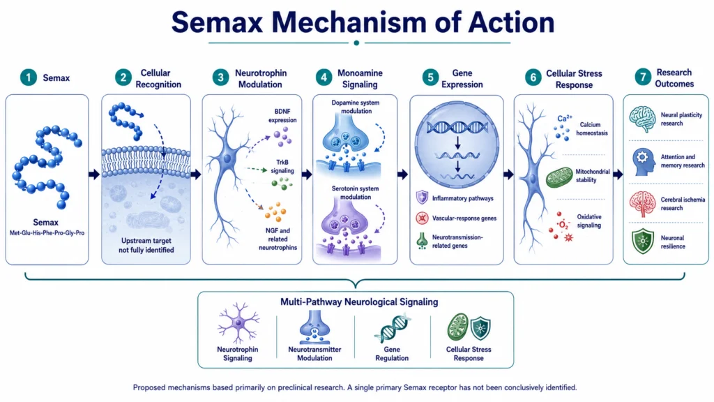
The phrase “mechanism of action” refers to the biological processes through which a molecule produces its effects. In the case of Semax, research suggests that it influences neurotrophin signaling, monoamine neurotransmitter systems, injury-responsive gene expression, and cellular responses to neurological stress.¹–⁹
Semax is best understood as a multi-pathway regulatory peptide. Unlike compounds with one clearly established receptor target, Semax does not have a single confirmed receptor-and-signaling cascade that fully explains its activity.
Instead, animal and cell studies suggest that its effects may emerge from several interconnected processes involving brain-derived neurotrophic factor, or BDNF; its receptor TrkB; other neurotrophins; dopamine and serotonin systems; inflammatory signaling; and neuronal stress resistance.
As discussed in What Is Semax?, Semax is derived from a short ACTH sequence but was developed without the primary corticotropic activity of full-length ACTH.
Semax Mechanism of Action: The Science Explained
Semax’s proposed mechanism can be summarized as:
- Region-specific peptide binding and upstream signaling
- Modulation of the BDNF/TrkB system
- Changes in broader neurotrophin gene expression
- Regulation of dopamine and serotonin systems
- Modification of inflammatory and neurotransmission-related gene expression
- Support of cellular responses to excitotoxic and ischemic stress
These processes should not be viewed as one proven linear cascade. Current evidence suggests that Semax produces time-dependent and region-specific effects across several neurological pathways.
1. Region-Specific Peptide Binding and Upstream Signaling
The first step in Semax’s mechanism remains one of the least clearly defined.
A study using rat basal forebrain tissue reported specific Semax binding, suggesting that the peptide interacts with particular cellular binding sites rather than acting only through nonspecific chemical effects.²
However, the molecular identity of those binding sites was not established. Semax has therefore not been assigned one confirmed primary receptor comparable to the GHRH receptor for CJC-1295.
This distinction is important because many of the pathways associated with Semax—such as BDNF expression or inflammatory gene regulation—may be downstream responses rather than evidence that Semax directly binds the proteins involved.
Why this matters:
The available research supports specific biological recognition of Semax, but the initial receptor or signaling target has not been conclusively identified. Its mechanism should therefore be described as proposed and multi-pathway rather than as one settled receptor cascade.
2. BDNF and TrkB Signaling
The most frequently studied Semax pathway involves brain-derived neurotrophic factor and its receptor TrkB.
BDNF is a neurotrophin involved in neuronal development, synaptic adaptation, and cellular responses to neurological stress. BDNF activates TrkB, a receptor expressed throughout the nervous system.
In rodent studies, intranasal Semax altered BDNF expression and the activation or expression of TrkB in the hippocampus.¹ Another study reported specific Semax binding and increased BDNF protein levels in the rat basal forebrain.²
The available findings support a model in which Semax influences the production and signaling environment of BDNF. They do not demonstrate that Semax binds directly to TrkB or functions as a conventional TrkB agonist.
A simplified version of the proposed pathway is:
Semax exposure → increased or modified BDNF expression → BDNF-mediated TrkB signaling → downstream changes in neuronal adaptation
The timing and direction of these changes may vary by brain region and experimental conditions.
Why this matters:
BDNF/TrkB signaling offers one biological explanation for Semax’s relevance to research on neural plasticity, learning, and neurological recovery. However, BDNF is best understood as a downstream pathway influenced by Semax—not as a confirmed direct receptor target.
3. Broader Neurotrophin Gene Expression
Semax research extends beyond BDNF alone.
In a rat model of cerebral ischemia, researchers examined the expression of multiple neurotrophins and their receptors after Semax exposure. The study reported time-dependent changes involving:
- BDNF
- Nerve growth factor, or NGF
- Neurotrophin-3, or NT-3
- TrkA
- TrkB
- TrkC
Semax enhanced the transcription of different neurotrophin and receptor genes at different stages after cerebral artery occlusion.³
This suggests that Semax may affect a broader neurotrophic response rather than activating one isolated growth-factor pathway.
The results also varied according to timing and brain region. This indicates that Semax may influence the nervous system’s adaptive response differently depending on the condition being studied.
Why this matters:
The broader neurotrophin findings help explain why Semax cannot be reduced to a “BDNF peptide.” BDNF is a major research focus, but Semax appears to influence a wider network of neurotrophins and receptors.
4. Dopamine and Serotonin System Modulation
Semax has also been studied for its effects on dopaminergic and serotonergic signaling.
In rodents, Semax altered neurochemical measurements associated with dopamine and serotonin in several brain regions.⁴ The effects were region-specific and did not amount to a simple, uniform increase in both neurotransmitters throughout the brain.
Dopamine and serotonin systems participate in processes such as:
- Attention
- Motivation
- Learning
- Behavioral adaptation
- Stress responses
- Mood regulation
Changes in these systems are one reason Semax is frequently discussed in cognitive research. However, the available studies do not show that Semax directly binds dopamine or serotonin receptors.
Instead, Semax appears to modify the activity or metabolism of monoamine systems through an indirect and incompletely understood process.
Why this matters:
Monoamine modulation provides a possible link between Semax’s molecular effects and its use in behavioral and cognitive research. The evidence remains preclinical and should not be simplified into claims that Semax reliably “raises dopamine” or “increases serotonin” in humans.
5. Inflammatory and Neurotransmission-Related Gene Expression
Some of the most detailed Semax research has examined how it changes gene expression after experimental cerebral ischemia.
A genome-wide study in rats found that Semax affected genes associated with immune responses and vascular function after focal brain ischemia.⁵ Later transcriptomic research reported that ischemia-reperfusion activated inflammatory genes while suppressing genes related to neurotransmission. Semax shifted portions of this pattern in the opposite direction.⁶
In that model, Semax was associated with:
- Reduced expression of several inflammation-related genes
- Increased expression of genes connected to neurotransmission
- Partial correction of gene-expression patterns disrupted by ischemia
- Changes in immune- and vascular-related signaling
Protein-expression research has also examined markers involved in inflammation, cell death, stress responses, and recovery, including MMP-9, JNK, c-Fos, and CREB.⁷
The cleanest interpretation is not that Semax blocks one inflammatory molecule. Instead, it may modify broader transcriptional programs activated during neurological injury.
Why this matters:
This gene-regulatory profile may help explain why Semax is studied in models of neurological stress and cerebral ischemia. It also demonstrates that some of its proposed effects involve neuroimmune and vascular pathways, not neurons alone.
6. Cellular Responses to Excitotoxic and Ischemic Stress
Semax has also been investigated at the level of cellular stress resistance.
Excessive glutamate exposure can disrupt intracellular calcium regulation, impair mitochondrial membrane potential, and contribute to neuronal injury. In cultured cerebellar neurons exposed to glutamate toxicity, Semax and its Pro-Gly-Pro fragment delayed calcium dysregulation and loss of mitochondrial potential. They were also associated with improved neuronal survival in that experimental model.⁸
A separate rat study examined nitric oxide and lipid-peroxidation changes following incomplete global cerebral ischemia. Semax reduced the ischemia-associated increase in nitric oxide generation and affected markers of oxidative damage.⁹
These findings suggest that Semax’s proposed neuroprotective profile may involve several cellular processes:
- Calcium homeostasis
- Mitochondrial stability
- Oxidative signaling
- Responses to excessive glutamate
- Cellular adaptation to reduced blood flow
These studies were conducted in cultured cells or animal ischemia models. They do not establish that Semax prevents neuronal damage in humans.
Why this matters:
Neurotrophin and gene-expression changes may be only part of Semax’s research profile. Its observed effects on calcium regulation, mitochondrial function, and oxidative pathways suggest that it may also modify how cells respond to acute neurological stress.
Additional Proposed Enzyme Effects
The strongest direct human data on CJC-1295 involve the DAC-modified lonSemax has also been studied as a potential inhibitor of enzymes that degrade enkephalins and other regulatory peptides.
An in vitro study using human serum found that Semax inhibited enkephalin-degrading enzyme activity in a concentration-dependent manner.¹⁰ The researchers proposed that slowing the degradation of endogenous regulatory peptides could contribute to Semax’s biological activity.
However, this finding has not been established as a central in vivo mechanism. The concentrations required, the specific enzymes involved, and the clinical relevance remain uncertain.
This pathway is therefore best treated as a secondary mechanistic hypothesis rather than a confirmed explanation of Semax’s neurological effects.
Semax’s Multi-Pathway Neurological Profile
Unlike CJC-1295, which follows a defined endocrine pathway from GHRH receptor activation to GH and IGF-1 signaling, Semax appears to act through a network of neurological responses.
| Proposed pathway | Main experimental observation | Evidence base |
| Initial peptide recognition | Specific binding reported in rat basal forebrain tissue | Preclinical tissue research |
| BDNF/TrkB signaling | Changes in BDNF protein, gene expression, and TrkB activity or expression | Rodent studies |
| Broader neurotrophin signaling | Changes in BDNF, NGF, NT-3, and Trk receptor transcription | Rodent ischemia models |
| Monoamine systems | Region-specific changes in dopamine- and serotonin-related measurements | Rodent studies |
| Neuroimmune regulation | Modification of inflammatory, vascular, and neurotransmission-related genes | Rodent ischemia models |
| Cellular stress response | Changes in calcium regulation, mitochondrial potential, nitric oxide, and neuronal survival | Cell and animal studies |
| Peptidase inhibition | Reduced enkephalin-degrading enzyme activity | Human-serum laboratory study |
A simplified research model is:
Semax recognition → neurotrophin and neurotransmitter modulation → changes in gene expression and cellular stress responses
This model is useful for organizing the evidence, but it should not be interpreted as a fully confirmed sequence of events.
Limitations: What Do Studies Say?
The most important limitation is that Semax does not have one conclusively identified primary receptor.
The research demonstrates changes in several biological pathways, but it is not always clear which changes are direct and which are secondary consequences of earlier signaling events.
Other limitations include:
- Most mechanistic studies use rats, cultured neurons, or experimental cerebral-ischemia models.
- Findings can differ according to brain region, dose, timing, and experimental conditions.
- Increased BDNF or altered gene expression does not automatically establish a meaningful behavioral or clinical effect.
- Semax has not been shown to bind directly to TrkB, dopamine receptors, or serotonin receptors.
- The Pro-Gly-Pro region and peptide breakdown products may contribute to some observed effects.
- Human studies have not fully validated the multi-step mechanisms proposed from preclinical research.
The clean scientific distinction is:
Well-supported preclinical observation: Semax alters neurotrophin, monoamine, gene-expression, and cellular-stress markers in experimental models.
Not yet established: One primary receptor, one universal signaling cascade, or a fully validated human mechanism of action.
Conclusion
Semax appears to work through several interconnected neurological pathways rather than one confirmed receptor mechanism.
The most studied pathway involves modulation of BDNF and TrkB signaling. Semax has also been associated with changes in other neurotrophins, dopamine and serotonin systems, inflammatory and neurotransmission-related gene expression, calcium regulation, mitochondrial stability, and oxidative responses.¹–⁹
Some studies have reported specific Semax binding, but the responsible receptor or binding protein has not been conclusively identified. Semax should therefore not be described as a direct BDNF, TrkB, dopamine, or serotonin receptor agonist.
The most accurate explanation is that Semax is a multi-pathway regulatory peptide whose observed effects depend on the brain region, timing, and experimental model being studied.
FAQs About Semax Mechanism
How does Semax work?
Semax appears to influence several neurological pathways, including BDNF/TrkB signaling, broader neurotrophin expression, dopamine and serotonin systems, inflammatory gene regulation, and cellular responses to ischemic or excitotoxic stress.
What receptor does Semax bind to?
No single primary receptor has been conclusively established. Research has reported specific Semax binding in rat brain tissue, but the molecular identity of the binding site remains unclear.
Does Semax bind directly to TrkB?
Current research does not establish Semax as a direct TrkB agonist. Instead, animal studies suggest that Semax changes BDNF expression and the activation or expression of the BDNF receptor TrkB.
Is BDNF the main mechanism of Semax?
BDNF/TrkB is the most frequently studied Semax pathway, but it is unlikely to explain the peptide’s complete research profile. Semax has also been associated with other neurotrophins, neurotransmitter systems, immune signaling, and cellular stress responses.
Does Semax increase dopamine and serotonin?
Rodent studies have reported region-specific changes in dopaminergic and serotonergic systems. The findings do not support a simple conclusion that Semax uniformly increases dopamine or serotonin throughout the brain or reliably produces the same effects in humans.
Does Semax reduce inflammation?
In rat cerebral-ischemia models, Semax modified the expression of several inflammation-related genes. These findings are model-specific and do not establish Semax as a general anti-inflammatory treatment.
How could Semax affect neuronal survival?
Cell and animal research suggests that Semax may influence neurotrophin signaling, calcium regulation, mitochondrial membrane stability, oxidative pathways, and injury-responsive gene expression. These processes may contribute to neuronal survival in experimental stress models.
Does the Pro-Gly-Pro portion of Semax contribute to its mechanism?
Possibly. Pro-Gly-Pro produced some overlapping effects with Semax in neurotrophin and cellular-stress studies. This suggests that the PGP portion or related peptide fragments may contribute to some of Semax’s biological activity.
Does Semax inhibit enkephalin breakdown?
One laboratory study found that Semax inhibited enkephalin-degrading enzyme activity in human serum. The importance of this finding in living organisms and at relevant exposures remains uncertain.
Is the Semax mechanism proven in humans?
No. Most detailed mechanistic evidence comes from animal and cell studies. Limited human research has not established one complete or independently replicated mechanism of action.
Related Articles
References
- Dolotov OV, Karpenko EA, Inozemtseva LS, et al. Semax, an analog of ACTH (4-10) with cognitive effects, regulates BDNF and TrkB expression in the rat hippocampus. Brain Research. 2006;1117(1):54–60. https://pubmed.ncbi.nlm.nih.gov/16996037/
- Dolotov OV, Karpenko EA, Seredenina TS, et al. Semax, an analogue of adrenocorticotropin (4-10), binds specifically and increases levels of brain-derived neurotrophic factor protein in rat basal forebrain. Journal of Neurochemistry. 2006;97(Suppl 1):82–86. https://pubmed.ncbi.nlm.nih.gov/16635254/
- Dmitrieva VG, Povarova OV, Skvortsova VI, Limborska SA, Myasoedov NF, Dergunova LV. Semax and Pro-Gly-Pro activate the transcription of neurotrophins and their receptor genes after cerebral ischemia. Cellular and Molecular Neurobiology. 2010;30(1):71–79. https://pubmed.ncbi.nlm.nih.gov/19633950/
- Eremin KO, Kudrin VS, Saransaari P, et al. Semax, an ACTH (4-10) analogue with nootropic properties, activates dopaminergic and serotoninergic brain systems in rodents. Neurochemical Research. 2005;30(12):1493–1500. https://pubmed.ncbi.nlm.nih.gov/16362768/
- Medvedeva EV, Dmitrieva VG, Povarova OV, et al. The peptide Semax affects the expression of genes related to the immune and vascular systems in rat brain focal ischemia: genome-wide transcriptional analysis. BMC Genomics. 2014;15:228. https://pmc.ncbi.nlm.nih.gov/articles/PMC3987924/
- Filippenkov IB, Stavchansky VV, Denisova AE, et al. Novel insights into the protective properties of ACTH (4-7) PGP (Semax) peptide at the transcriptome level following cerebral ischaemia-reperfusion in rats. Genes. 2020;11(6):681. https://pmc.ncbi.nlm.nih.gov/articles/PMC7350263/
- Sudarkina OY, Filippenkov IB, Stavchansky VV, et al. Brain protein expression profile confirms the protective effect of the ACTH (4-7) PGP peptide Semax in a rat model of cerebral ischemia-reperfusion. International Journal of Molecular Sciences. 2021;22(12):6179. https://pmc.ncbi.nlm.nih.gov/articles/PMC8226508/
- Storozhevykh TP, Tukhbatova GR, Senilova YE, Pinelis VG, Andreeva LA, Myasoedov NF. Effects of Semax and its Pro-Gly-Pro fragment on calcium homeostasis of neurons and their survival under conditions of glutamate toxicity. Bulletin of Experimental Biology and Medicine. 2007;143(5):601–604. https://pubmed.ncbi.nlm.nih.gov/18239779/
- Bashkatova VG, Koshelev VB, Fadyukova OE, et al. Novel synthetic analogue of ACTH 4-10 (Semax) but not glycine prevents the enhanced nitric oxide generation in cerebral cortex of rats with incomplete global ischemia. Brain Research. 2001;894(1):145–149. https://pubmed.ncbi.nlm.nih.gov/11245825/
- Kost NV, Sokolov OY, Gabaeva MV, et al. Semax and Selank inhibit the enkephalin-degrading enzymes from human serum. Russian Journal of Bioorganic Chemistry. 2001;27(3):180–183. https://pubmed.ncbi.nlm.nih.gov/11443939/
