The mitochondrial peptide linking energy, exercise, and healthy aging
Disclaimer: Information provided is for research and educational purposes only. MOTS-c is not approved by the FDA or any regulatory agency for therapeutic or cosmetic use.
Introduction:
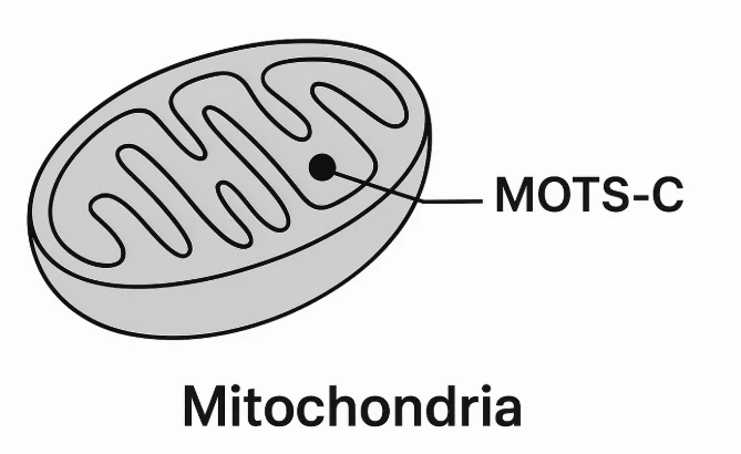
MOTS-c (short for mitochondrial open reading frame of the 12S rRNA-c) is a 16-amino-acid peptide encoded by mitochondrial DNA. Discovered in 2015, it helps cells adapt to metabolic stress by optimizing energy use, improving insulin sensitivity, and maintaining mitochondria function.¹
Unlike most peptides (encoded in nuclear DNA), MOTS-c originates inside mitochondria and can even move to the nucleus to influence gene expression—an unusual form of mitochondria-to-nucleus signaling.²
Early research suggests MOTS-C may play a role in energy regulation, insulin sensitivity, exercise performance, and healthy aging. ³
MOTS-C is encoded by mitochondrial DNA, unlike most peptides which originate from nuclear DNA
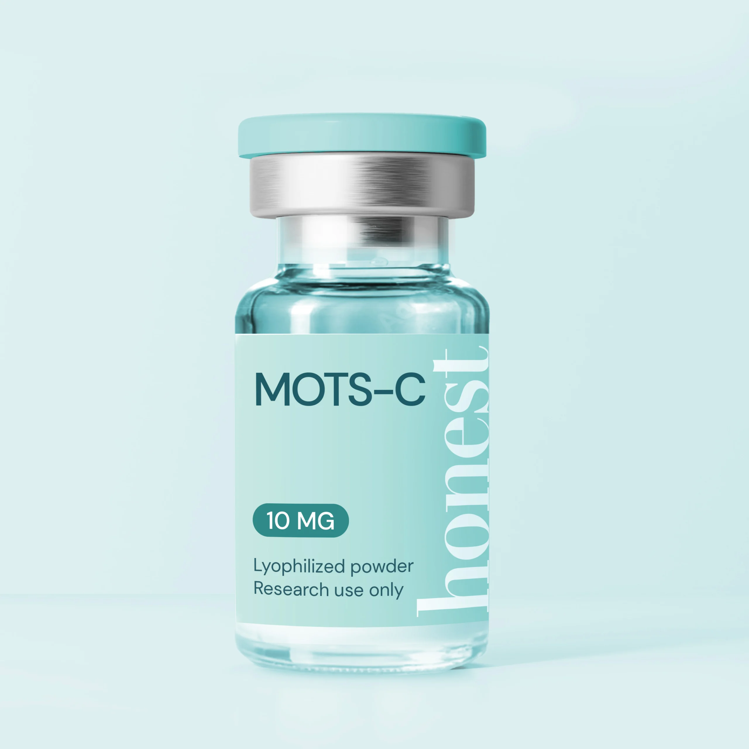
MOTS-c Fast Facts
Source: Encoded within the mitochondrial 12S rRNA gene (mtDNA).¹
Length: 16 amino acids.
Core actions: Activates AMPK, enhances glucose uptake, supports mitochondrial quality control, and regulates nuclear genes under stress.¹²
Where it shows up: Skeletal muscle (notably during/after exercise), liver, and circulation.¹³
Research interests: Insulin resistance and metabolic health, exercise physiology, aging biology.¹³⁴
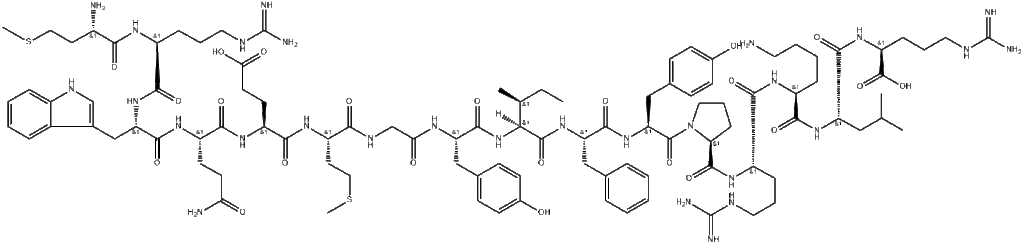
Chemical Structure
MOTS-c is a 16-amino-acid peptide encoded by the mitochondrial 12S rRNA gene. Its short, stable sequence allows it to act rapidly during energy stress, such as exercise or fasting, when cells need to rebalance fuel use and protect mitochondria.
MOTS-c Chemical Structure
How MOTS-c Works (In Brief)
When the body experiences energy stress — such as during exercise, fasting, or low nutrient availability — MOTS-c levels rise. It activates AMPK, the cell’s energy sensor, shifting metabolism toward fat oxidation and improved glucose control.¹
Uniquely, MOTS-c can also enter the nucleus and activate protective stress-response genes, demonstrating that mitochondria don’t just generate energy — they can also direct cellular adaptation.²
Discovery & Research Milestones
Direct links to all these studies can be found at the bottom of the article
Year
Study & Source
Key Finding
2015
Lee et al.¹
Discovery of MOTS-c as a mitochondrial-encoded peptide that improves insulin sensitivity and protects against diet-induced obesity.
2016
Lee et al.¹²
Follow-up work framed MOTS-c as a master regulator of cellular metabolism through AMPK activation and energy-balance pathways
2018
Kim et al.²
Found that MOTS-c can enter the nucleus to turn on stress-response genes.
2019
Benayoun & Lee¹¹
Framed MOTS-c as a messenger between mitochondria and the nucleus.
2020
D’Souza et al⁴
Showed MOTS-c levels decline with age in blood but rise in muscle.
2021
Reynolds et al.³
Identified MOTS-c as exercise-induced, improving endurance and strength.
2021
Yu et al.⁵
Demonstrated MOTS-c supports mitochondrial health via AMPK activation.
2021
Yang et al.¹⁰
Found MOTS-c and exercise work synergistically to enhance glucose control.
2022-2023
Mohtashami 2022⁸; Wan 2023⁶; Zheng 2023⁷; Kong 2023⁷
Recent reviews position MOTS-c as a promising link between metabolism, aging, and exercise physiology.
A Closer Look: What Research Shows
Metabolism & insulin sensitivity In mouse models of diet-induced obesity, MOTS-c improves glucose tolerance and prevents insulin resistance, partly via AMPK.¹
Exercise and Endurance MOTS-c rises with exercise; giving MOTS-c in animal studies boosts endurance and supports muscle adaptation across ages.³
Mitochondrial health In human stem-cell models, MOTS-c improves mitochondrial homeostasis (in turn linked to higher energy levels) and reduces ROS while activating AMPK and rebalancing mTORC1.⁵
Aging signals In humans, plasma MOTS-c tends to decline with age, while muscle MOTS-c can increase—hinting at a compensatory response.⁴
Summary
MOTS-c is a mitochondrial-encoded peptide that helps cells balance energy use, resist metabolic stress, and coordinate mitochondrial and nuclear responses. By activating AMPK, enhancing glucose control, and influencing gene expression, MOTS-c bridges the gap between energy metabolism, exercise physiology, and aging biology.While evidence is strongest in animal and cellular models, early human findings make MOTS-c a leading target in metabolism and longevity research.¹–¹²
FAQs About MOTS-C
What does MOTS-C stand for?
MOTS-C stands for “Mitochondrial Open Reading Frame of the 12S rRNA-c.”
Is MOTS-C naturally found in humans?
Yes, it is naturally expressed in skeletal muscle and circulates in the blood, especially during exercise or stress.
Does MOTS-C improve weight loss?
Animal studies suggest improved insulin sensitivity and fat metabolism, but human data is still limited.
Is MOTS-C legal to use as a supplement?
No, MOTS-C is research-only and not FDA-approved.
Related Articles
References
Lee C, Zeng J, Drew BG, et al. The mitochondrial-derived peptide MOTS-c promotes metabolic homeostasis and reduces obesity and insulin resistance.Cell Metab. 2015;21(3):443-454. https://pubmed.ncbi.nlm.nih.gov/25738459
Kim KH, Son JM, Benayoun BA, Lee C. The mitochondrial-encoded peptide MOTS-c translocates to the nucleus to regulate nuclear gene expression in response to metabolic stress.Cell Metab. 2018;28(4):516-524.e7. https://pubmed.ncbi.nlm.nih.gov/29983246/
Reynolds JC, Mazzucco AE, Chen CY, et al. MOTS-c is an exercise-induced mitochondrial-encoded regulator of age-dependent physical decline and muscle homeostasis.Nat Commun. 2021;12:470. https://pubmed.ncbi.nlm.nih.gov/33473109
D’Souza RF, Woodhead JST, Zeng N, et al. Increased expression of the mitochondrial derived peptide, MOTS-c, in skeletal muscle of healthy aging men is associated with myofiber composition.Aging (Albany NY). 2020;12(6):5244-5265. https://pubmed.ncbi.nlm.nih.gov/32182209
Yu WD, Wang HB, Teng L, et al. MOTS-c promotes mitochondrial homeostasis in human placenta-derived mesenchymal stem cells via AMPK activation and mTORC1 inhibition.J Tissue Eng Regen Med. 2021;15(3):254-267. https://pubmed.ncbi.nlm.nih.gov/33639272
Yang B, Yu Q, Chang B, et al. MOTS-c interacts synergistically with exercise to attenuate insulin resistance and enhance glucose metabolism via AMPK–PGC-1α signaling (mouse).Biochim Biophys Acta Mol Basis Dis. 2021;1867(6):166126. https://pubmed.ncbi.nlm.nih.gov/33722744
The melanocortin peptide studied for tanning and sexual function
Disclaimer: Information provided is for research and educational purposes only. Melanotan-II is not approved by the FDA or EMA for human or cosmetic use.
Melanotan-II (MT-II), often called the “tanning peptide,” is a synthetic analog of the natural α-melanocyte-stimulating hormone (α-MSH). It was developed in the 1980s to explore sunless tanning and skin-protection effects.¹ As a melanocortin receptor agonist, Melanotan-II promotes skin pigmentation and has been studied for its influence on sexual function, energy balance, and UV protection.¹ ² ³ ⁴ ⁸ ⁹
Design: Cyclic heptapeptide (lactam bridge) engineered for potency/stability
Primary targets: MC1R (pigmentation), central MC3R/MC4R (sexual function, energy balance).⁸
Most studied effects: Increased pigmentation; erectogenic response in ED cohorts; nausea/yawning common.¹
Melanotan-2 Chemical Structure
What exactly does MT-II do?
MT-II stimulates melanogenesis via MC1R on melanocytes → darker pigmentation following short subcutaneous dosing.¹
In the CNS, MT-II can trigger erections without sexual stimulation, pointing to central melanocortin circuitry (hypothalamic and spinal sites).²
Discovery & Research Milestones
Originated at the University of Arizona in the 1980s, where researchers were exploring sunless tanning agents.¹
Built as a short, stable analog of α-MSH, improving its half-life and activity compared to the natural hormone.²
Early studies tested Melanotan II in skin pigmentation disorders and sexual dysfunction, but it never achieved regulatory approval.³
Year
Study & Source
Key Finding
1996
Dorr RT et al.
Visible tanning after low-dose MT-II; common side effects include nausea & yawning. Recommended 0.025 mg/kg.
1998
Wessells H et al.
First studies on use for erectile dysfunction. MT-2 induces erections without erotic stimuli
2000-2003
Wessells H et al.
Mechanism established: central melanocortin pathways (hypothalamus and spinal cord) mediate MT-II-induced erection.
2003-2004
PT-141 program
MT-II signal translated into PT-141/bremelanotide; early RCTs show dose-dependent erectile activity and acceptable tolerance.
A Closer Look: What the Research Shows
Skin Pigmentation (Tanning) A single-blind, alternating-day phase-I trial found visible tanning after five low doses and recommended 0.025 mg/kg for future studies; common AEs were nausea, yawning, somnolence.
Erectile physiology A double-blind, placebo-controlled crossover study showed robust erections (average of 38 min of >80% rigidity) with 0.025 mg/kg MT-II; nausea/yawning increased vs. placebo.
Development of PT-141 The erectogenic signal from MT-II led to development of PT-141/bremelanotide (a related melanocortin agonist)
Research Applications
Pigmentation disorders: Investigated for use in vitiligo and erythropoietic protoporphyria.²
Skin protection: Studied for potential reduction of UV-related skin damage.³
Sexual function: Explored as a treatment for erectile dysfunction and female sexual arousal disorder. ³
Regulatory Status
MT-II: Not approved for tanning or sexual function
Afamelanotide (MT-I) is approved for EPP (EU 2014; US 2019); long-term safety/PK are reviewed separately.
Summary
Melanotan-2 is a potent melanocortin agonist that darkens skin (via MC1R) and triggers erections by acting on central melanocortin pathways. Controlled early trials defined doses and common AEs; the erectogenic signal catalyzed development of bremelanotide.
FAQs About Melanotan II
Is Melanotan II FDA-approved?
No, it is not approved by the FDA or EMA and is considered a research chemical.
What does Melanotan II do?
It increases melanin production in the skin by activating melanocortin receptors, leading to darker pigmentation.
What was Melanotan II originally developed for?
It was developed as a tanning agent and studied for pigmentation disorders and sexual dysfunction.
Dorr RT, Lines R, Levine N, et al.Evaluation of melanotan-II, a superpotent cyclic melanotropic peptide in a pilot phase I clinical study.Life Sci. 1996;58(20):1777-1784. https://pubmed.ncbi.nlm.nih.gov/8637402/
Wessells H, Fuciarelli K, Hansen J, et al.Synthetic melanotropic peptide initiates erections in men with psychogenic erectile dysfunction: double-blind, placebo-controlled crossover study.J Urol. 1998;160(2):389-393. https://pubmed.ncbi.nlm.nih.gov/9679884/
Vemulapalli R, Kurosawa M, Killinger J, et al.Activation of central melanocortin receptors by melanotan-II (Ac-Nle-c[Asp-His-D-Phe-Arg-Trp-Lys]-NH₂) results in penile erection in rats.Neuroscience. 2001;106(3):547-552. https://pubmed.ncbi.nlm.nih.gov/11591452/
Wessells H, Hruby V J, Hackett J, et al.Central melanocortin pathways mediate the erectogenic effects of melanotan-II in animal models.J Urol. 2003;170(6 Pt 1):S. (Abstract and mechanistic report.) https://pubmed.ncbi.nlm.nih.gov/14665870/
Rosen R C, Diamond L E, Earle D C, Shadiack A M, Molinoff P B.Evaluation of the safety, pharmacokinetics and pharmacodynamics of intranasal PT-141, a melanocortin receptor agonist, in healthy males and patients with erectile dysfunction.J Sex Med. 2004;1(3):193-202. https://pubmed.ncbi.nlm.nih.gov/14999221/
Diamond L E, Earle D C, Garcia W D, Spana C.Double-blind, placebo-controlled evaluation of the safety, efficacy and dose response of subcutaneous PT-141 in men with erectile dysfunction.Int J Impot Res. 2004;16(4):353-360. https://pubmed.ncbi.nlm.nih.gov/14963471/
Hadley M E.Discovery that a melanocortin regulates sexual functions in male and female humans.Peptides. 2005;26(10):1687-1691. https://pubmed.ncbi.nlm.nih.gov/15996790/
Hjuler K F, Brøgger-Mikkelsen M, Pedersen S, et al.Melanoma associated with the use of melanotan-II.Acta Derm Venereol. 2014;94(1):115-116. https://pubmed.ncbi.nlm.nih.gov/24355990/
Minder E I, Schneider-Yaniv L, Schneider-Yaniv Z.Pharmacokinetics and pharmacodynamics of afamelanotide, a melanocortin 1 receptor agonist, in humans.Clin Pharmacokinet. 2017;56(8):815-826. https://pubmed.ncbi.nlm.nih.gov/28063031/
The hGH Fragment Studied for Fat Metabolism and Weight Regulation
Disclaimer: Information provided is for research and educational purposes only. AOD-9604 is not approved by the FDA for therapeutic or cosmetic use.
Introduction:
AOD-9604 is a synthetic peptide fragment of human growth hormone (hGH 177–191) engineered to replicate hGH’s fat-metabolism effects without altering IGF-1 or glucose levels.¹ In preclinical research, it promoted lipolysis (fat breakdown) and reduced lipogenesis (fat storage) through receptor-independent mechanisms.² ³
AOD-9604 Fast Facts
Class: Synthetic hGH fragment analog (Tyr-hGH 177–191)¹
Intended purpose: Stimulate fat metabolism while avoiding hGH’s growth or glucose effects²
Distinct from hGH: Does not activate the growth hormone receptor or raise IGF-1² ⁴
Development stage: Completed Phase II trials; discontinued after lack of clinical efficacy⁶ ⁹
Chemical Structure
AOD-9604 is a 15-amino-acid fragment of human growth hormone. It was engineered isolate hGH’s lipolytic (fat-breaking) properties without activating the growth hormone receptor or increasing IGF-1.² The design allows it to mimic hGH’s metabolic domain in a shorter, more stable form.
How AOD-9604 Works (in Brief)
AOD-9604 was engineered from the C-terminal segment of human growth hormone (amino acids 177–191) — the portion thought to influence fat metabolism.¹ ²
In preclinical models, this fragment supported fat oxidation and lipid mobilization without the broader hormonal effects of full-length hGH.³ ⁴
Because it does not activate the traditional hGH receptor, its fat-metabolism effects are considered indirect and receptor-independent.²
Discovery & Research Milestones
Originally developed in the 1990s as a potential anti-obesity drug. While it showed anti-obesity signal⁶ and advanced into clinical trials, it failed to show sufficient weight-loss effects to support commercialization.⁸
Year
Study & Source
Key Finding
2000 2001
Heffernan MA et al¹ ² ³
AOD-9604 found to increase fat oxidation and reduce weight gain.
2000
Ng FM et al.⁴
Demonstrated metabolic benefits without hGH-induced insulin resistance.
12-week trial showed modest signal; 24-week trial failed. Development stopped.
2013
Stier H et al. ⁵
Summarized human safety data—well tolerated, no antibodies detected.
AOD-9604 vs HGH Fragment 176–191: What’s the Difference?
Since AOD-9604 is often described as an HGH fragment, it’s easy to confuse the two. In reality:
HGH Fragment 176–191 is the natural sequence of amino acids (176 to 191) at the C-terminal end of human growth hormone (HGH).
AOD-9604 is a modified version of this fragment, designed to enhance stability and activity in fat metabolism. ¹²
Takeaway: AOD-9604 should be thought of as an optimized, research-developed analog of HGH fragment 176–191
Feature
HGH Fragment 176–191
AOD-9604
Origin
Direct amino acid sequence from HGH
Synthetic analog of fragment 176–191
Primary Action
Stimulates lipolysis (fat breakdown)
Stimulates lipolysis + inhibits lipogenesis
Stability
Shorter half-life, limited clinical exploration
Designed to be more stable and suitable for trials
Clinical Development
Investigated mainly in preclinical studies
Entered Phase I/II obesity trials
Takeaway
AOD-9604 should be thought of as an optimized, research-developed analog of HGH fragment 176–191 — not an entirely separate peptide, but not exactly the same either. This distinction helps explain why it progressed into formal obesity trials, whereas the raw fragment itself did not.
Regulatory Status
AOD-9604 is not FDA-approved for any medical use and is listed by the FDA as a bulk drug substance that may present safety risks.⁸ Clinical development was halted after Phase IIb trials failed to meet efficacy endpoints.⁹
Summary
AOD-9604 is a synthetic analog of the HGH 177–191 fragment developed for fat-metabolism research. Preclinical studies showed lipolytic and anti-lipogenic effects, but human trials failed to demonstrate significant weight-loss outcomes. It remains available for research use only.
FAQs About AOD-9604
What is AOD-9604?
AOD-9604 is a synthetic analog of the HGH fragment 176–191, studied for fat metabolism and weight regulation.
Is AOD-9604 the same as HGH fragment 176–191?
Not exactly — AOD-9604 is a modified version of the natural fragment, designed to be more stable and active.
Does AOD-9604 build muscle like HGH?
No, it does not stimulate IGF-1 or muscle growth — its effects are limited to fat metabolism pathways.
Is AOD-9604 approved for medical use?
No, AOD-9604 is not FDA-approved and is available only for research purposes.
Heffernan MA, et al. Effects of oral administration of a synthetic fragment of human growth hormone on lipid metabolism in rodents and in human adipose tissue. Am J Physiol Endocrinol Metab. https://pubmed.ncbi.nlm.nih.gov/10950816/
Heffernan MA, et al. Increase of fat oxidation and weight loss in obese mice by a fragment of human growth hormone. Obes Res. 2001;9(5):341–347. https://pubmed.ncbi.nlm.nih.gov/11673763/
Heffernan M, et al. The effects of human GH and its lipolytic fragment (AOD9604) on lipid metabolism in mice. Endocrinology. 2001;142(12):5182–5189. https://pubmed.ncbi.nlm.nih.gov/11713213/
Ng FM, et al. Metabolic studies of a synthetic lipolytic domain (AOD9604) of human growth hormone in vitro and in vivo. Ann N Y Acad Sci. 2000;921:262–272. https://pubmed.ncbi.nlm.nih.gov/11146367/
The Neuropeptide Studied for Sleep, Stress & Hormonal Regulation
Disclaimer: Information provided is for research and educational purposes only. DSIP is not approved by the FDA or any regulatory agency for therapeutic or cosmetic use.
Introduction:
Delta Sleep-Inducing Peptide (DSIP) is a neuropeptide composed of nine amino acids, first isolated in 1977 from rabbit brain extracts associated with slow-wave (delta) sleep.¹ ² ³ Initially characterized for its ability to promote deep, restorative sleep, DSIP was later found to also influence endocrine rhythms, stress adaptation, and neuronal excitability.⁴ ⁵ Despite decades of research and significant physiological effects, DSIP’s exact mechanism remains unresolved. ⁶ ⁷
DSIP Fast Facts
Property
Details
Full Name
Delta Sleep-Inducing Peptide
Sequence
Trp–Ala–Gly–Gly–Asp–Ala–Ser–Gly–Glu
Classification
Neuropeptide, sleep-related peptide
Discovered
1977 – Schoenenberger & Monnier, University of Basel¹ ²
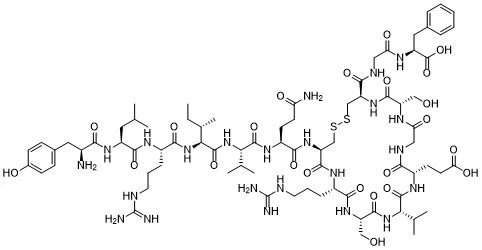
DSIP is a nonapeptide (nine-amino-acid chain) with the sequence Trp-Ala-Gly-Gly-Asp-Ala-Ser-Gly-Glu.¹ It was first isolated from rabbit hypothalamic extracts and later synthesized for comparative studies confirming identical biological activity.²
Unlike many peptides produced in classic endocrine glands, DSIP’s precise biosynthetic origin remains uncertain. Some evidence suggests it may arise from a larger pituitary precursor protein processed into DSIP-like fragments.⁷
Its short, linear structure makes it relatively unstable in plasma, contributing to the difficulty in verifying its natural circulation and consistent effects.⁹
DSIP Chemical Structure
How to DSIP Works (In Brief)
DSIP is thought to promote slow-wave (delta) sleep by modulating thalamo-cortical activity rather than acting as a sedative.¹–³ It may also couple sleep and growth-hormone release through hypothalamic pathways involving GHRH and somatostatin.⁶ Studies suggest interactions with GABAergic, serotonergic, and opioid systems, which could explain its effects on stress and arousal.⁴ ⁵ ⁸ Although DSIP-like fragments have been detected in the pituitary, its true biosynthesis and receptor targets remain unresolved, leaving its mechanism partly mysterious despite decades of study.⁷ ⁹
What Research Shows
1.DSIP improves Sleep Regulation: DSIP administration in animal models increases delta-wave activity and promotes slow-wave (non-REM) sleep without acting as a sedative.¹ ² ³
2.DSIP releases Growth Hormone Studies indicate DSIP may stimulate growth-hormone secretion during sleep and modulate corticotropin and gonadotropin rhythms.⁶
3. DSIP may regulate mood and be neuroprotective: Experimental data suggest DSIP can attenuate stress-induced changes in corticosterone and may interact with GABAergic and opioid systems, implying a role in neuroprotection and mood regulation.⁴ ⁵ ⁸
Discovery and Scientific Background
DSIP was first isolated in 1974 by Monnier et al., who observed that injections of the peptide into rabbits promoted slow-wave sleep (delta sleep).¹ Over the decades, narrow sleep studies have expanded to broader neuroendocrine investigation.
Year
Study & Source
Key Finding
1977 – Discovery
Schoenenberger GA, Monnier M. Proc Natl Acad Sci USA ¹
Identified a brain-derived peptide inducing delta-wave sleep in rabbits.
1977 – Characterization
Monnier M et al. Experientia ²
Synthetic DSIP reproduced the natural peptide’s sleep-promoting effects.
1980 – Sleep in Rodents
Nagasaki H et al. Brain Res ³
Confirmed DSIP enhances slow-wave sleep in mice without sedation..
Reviewed DSIP’s potential roles in anesthesia, stress adaptation, and unresolved molecular mechanisms.
Summary
Delta Sleep-Inducing Peptide (DSIP) is a neuropeptide originally identified for its ability to promote deep, slow-wave sleep. Subsequent research expanded its profile to include endocrine modulation, stress adaptation, and possible neuroprotective functions. Though its endogenous origin and mechanism remain debated, DSIP remains a compelling subject in studies of sleep architecture and hypothalamic–pituitary regulation.¹–⁹
FAQs About DSIP
What does DSIP stand for?
Delta Sleep-Inducing Peptide
What is DSIP studied for?
Research focuses on its role in sleep cycles, stress modulation, and neuroendocrine regulation.
Is DSIP effective as a sleep aid?
Animal studies suggested sleep-inducing effects, but results in humans are inconsistent.
Is DSIP safe?
There is limited data on long-term safety. It is not approved for medical use.
Schoenenberger GA, Monnier M.Characterization of a delta-electroencephalogram (-sleep)-inducing peptide.Proc Natl Acad Sci U S A. 1977;74(3):1282–1286. https://pubmed.ncbi.nlm.nih.gov/265572/
Monnier M, Dudler L, Gächter R, et al.The delta sleep-inducing peptide (DSIP). Comparative properties of the original and synthetic nonapeptide.Experientia. 1977;33(4):548–552. https://pubmed.ncbi.nlm.nih.gov/862769/
Nagasaki H, Kitahama K, Valatx JL, Jouvet M.Sleep-promoting effect of SPS and DSIP in the mouse.Brain Res. 1980;192(1):276–280. https://pubmed.ncbi.nlm.nih.gov/7378787/
Iyer KS, Marks GA, Kastin AJ, McCann SM.Evidence for a role of delta sleep-inducing peptide in slow-wave sleep and sleep-related growth hormone release in the rat.Proc Natl Acad Sci U S A. 1988;85(10):3728–3732. https://pmc.ncbi.nlm.nih.gov/articles/PMC280272/
Bjartell A, Castro MG, Ekman R, Sundler F, Widerlöv E, Loh YP.Biosynthesis and processing of DSIP-like precursors in mouse anterior pituitary cultures.Eur J Biochem. 1990;190(1):131–137. https://pubmed.ncbi.nlm.nih.gov/2364941/
The Growth Hormone Secretagogue Studied for GH Release & Metabolic Research
Disclaimer: Information provided is for research and educational purposes only. Ipamorelin is not approved by the FDA or any regulatory agency for therapeutic use.
Introduction
Ipamorelin is a synthetic pentapeptide developed as a selective growth-hormone secretagogue (GHS). It binds to the growth-hormone secretagogue receptor (GHS-R1a) in the pituitary to trigger growth-hormone (GH) release while leaving other endocrine pathways—such as ACTH, cortisol, prolactin, and aldosterone—largely unaffected.¹ ² ³.
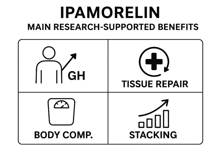
Known for its high receptor selectivity, predictable pharmacokinetics, and favorable safety profile, Ipamorelin has become a reference compound for studies on GH regulation, metabolism, and tissue repair.⁴ ⁵ ⁶
Discovered: Late 1990s – Novo Nordisk research team
Mechanism: Selective GHS-R1a agonist that stimulates GH release via ghrelin-like signaling
Key Features: Strong GH stimulation with minimal cortisol or prolactin response ¹ ² ³
Primary Research Areas: Growth hormone release, bone formation, metabolism, tissue repair ⁴ ⁵ ⁶ ⁷ ⁸ ⁹
Chemical Structure
Ipamorelin is a synthetic peptide – designed as a refined analog of earlier GHRPs – developed to isolate ghrelin’s growth hormone release without raising cortisol or prolactin. Its modified amino acids — including Aib and two D-residues — make it more resistant to breakdown and reduce unwanted hormonal effects.
Ipamorelin peptide structure and amino acid sequence
How Ipamorelin Works (in Brief)
Ipamorelin selectively activates GHS-R1a receptors on pituitary somatotroph cells, leading to dose-dependent GH release.¹ ² Unlike earlier secretagogues such as GHRP-6 or Hexarelin, Ipamorelin shows little to no stimulation of cortisol or ACTH, indicating high pathway specificity.³ ⁴ Animal and cell studies suggest downstream effects on bone formation, nitrogen balance, and adipose metabolism distinct from direct GH administration.⁵ ⁶ ⁷
Discovery & Research Milestones
Year
Study & Source
Key Finding
1998
Raun K et al.¹
Ipamorelin discovered as selective GHS with no ACTH or cortisol elevation
1999 2000 2001
Johansen PB et al.² Svensson J et al.⁴ Andersen NB et al.⁵
Studies showing how ipamorelin stimulates bone growth and bone health
1999
Gobburu JVS et al.³
First human PK/PD model; dose-proportional release with short half-life
2001
Lall S et al.⁶
Identified GH-independent adiposity effects of GHSs including ipamorelin.
2009
Aagaard NK et al.⁷
Showed modulation of hepatic nitrogen metabolism and urea synthesis in steroid-treated rats.
2014
Beck DE et al.⁸
Phase-2 clinical trial for postoperative ileus; ipamorelin was safe, with non-significant trend toward faster GI recovery.
Why is Ipamorelin Popular in Research?
Growth Hormone Stimulation: Promotes GH release without elevating other pituitary hormones ¹ ² ³
Bone and Tissue Repair: Improves bone formation and supports healing in animal models ⁴ ⁵ ⁶
Pharmacology & Safety: Short half-life, dose-proportional response, favorable safety data ³ ⁸ ⁹
Summary
Ipamorelin is a selective synthetic GHS that triggers growth-hormone release without altering other hormonal axes. Preclinical and early clinical studies show potential roles in growth regulation, bone metabolism, and recovery research. Its specificity, pharmacokinetic predictability, and safety record make it a cornerstone compound in the study of ghrelin-mimetic peptides.
FAQs About Ipamorelin
What is Ipamorelin?
Ipamorelin is a synthetic peptide and selective growth hormone secretagogue used in research to stimulate GH release with minimal side effects.
What does Ipamorelin do?
Ipamorelin selectively stimulates growth hormone secretion and is researched for muscle, body composition, and anti-aging effects.
How is Ipamorelin different from other peptides?
Unlike older GHS peptides, Ipamorelin has high GH selectivity and a lower risk of side effects. It is often compared or combined with CJC-1295.
Is Ipamorelin approved for human use?
No. Ipamorelin is for laboratory research use only and is not approved for human consumption or therapy.
Raun K, Hansen BS, Johansen NL, et al. Ipamorelin, a novel pentapeptide growth hormone secretagogue. Eur J Endocrinol. 1998;139(5):552–561. https://pubmed.ncbi.nlm.nih.gov/9849822/
Johansen PB, Nowak J, Skjaerbaek C, et al. Ipamorelin, a new growth-hormone-releasing peptide, induces longitudinal bone growth in rats. Growth Horm IGF Res. 1999;9(2):106–113. https://pubmed.ncbi.nlm.nih.gov/10611799/
Gobburu JVS, Agersø H, Jusko WJ, Ynddal L. Pharmacokinetic-pharmacodynamic modeling of ipamorelin, a growth hormone releasing peptide, in human volunteers. Pharm Res. 1999;16(9):1412–1416. https://pubmed.ncbi.nlm.nih.gov/10496658/
Andersen NB, Malmlöf K, Johansen PB, et al. The growth hormone secretagogue ipamorelin counteracts glucocorticoid-induced decrease in bone formation of adult rats. Growth Horm IGF Res. 2001;11(5):266–272. https://pubmed.ncbi.nlm.nih.gov/11735244/
Lall S, Tung LY, Ohlsson C, Jansson JO, Dickson SL. GH-independent stimulation of adiposity by GH secretagogues. Biochem Biophys Res Commun. 2001;280(1):132–138. https://pubmed.ncbi.nlm.nih.gov/11162489/
Aagaard NK, Johansen PB, Orskov H, et al.Growth hormone and growth hormone secretagogue effects on nitrogen balance and urea synthesis in steroid treated rats. Growth Horm IGF Res. 2009; 19(2): 154–160. https://pubmed.ncbi.nlm.nih.gov/19231263/
Beck DE, Christie NA, Davis B, et al. Prospective, randomized, controlled phase-2 study of ipamorelin for postoperative ileus. Int J Colorectal Dis. 2014; 29(12): 1529–1537. https://pubmed.ncbi.nlm.nih.gov/25331030/
The Growth Hormone–Releasing Peptide That Mimics the Body’s Own GHRH
Disclaimer: Information provided is for research and educational purposes only. Sermorelin is not approved by the FDA or any regulatory agency for therapeutic or cosmetic use.
Introduction
Sermorelin (GHRH 1-29 NH₂) is a synthetic peptide fragment of growth hormone–releasing hormone (GHRH) — the natural signal the brain uses to stimulate growth hormone (GH) secretion¹ ². Originally developed in the 1980s, it became one of the first tools scientists used to study how GH pulses are regulated and how that process changes with age.³⁴. Unlike recombinant GH therapy, which delivers the hormone directly, sermorelin encourages the pituitary to release GH naturally, preserving the body’s feedback loops and pulse rhythm.⁵⁶
Fast Facts About Sermorelin
Property
Details
Sequence
First 29 amino acids of endogenous GHRH
Peptide Class
Synthetic GHRH analog
First Developed
Early 1980s
Mechanism
Stimulates the pituitary gland to release GH in a physiologic, pulsatile pattern⁴⁸
Common Study Areas
Endocrine modulation, body composition, aging research
Chemical Structure
Sermorelin is a synthetic peptide containing the first 29 amino acids of natural human growth hormone releasing hormone (GHRH or somatocrinin). This fragment retains the full biological activity of the 44-amino-acid GHRH while offering greater stability and ease of synthesis due to its length.
Sermorelin chemical structure and amino acid sequence
Discovery & Research Milestones
GHRH itself was identified in 1982 from human pancreatic tumor extracts that caused acromegaly¹².The discovery of its sequence led to the synthesis of shorter, biologically active fragments, the most successful being GHRH (1-29) NH₂ — later named sermorelin.³ ⁷
By the mid-1980s, clinical studies confirmed that sermorelin robustly increased GH levels in healthy men and women without significantly altering other pituitary hormones.⁴ ⁵ ⁶ Researchers also observed that sermorelin produced natural GH pulses, resembling the body’s normal nighttime secretion pattern.⁸
Year
Study & Source
Key Finding
1982
Rivier J. et al.; Guillemin R. et al.¹ ²
Human growth hormone–releasing hormone (GHRH) isolated from pancreatic tumors, establishing the native GH-releasing pathway.
1983-1984
Thorner M.O. et al.; Barron J.L. et al.³ ⁴
Shorter fragments synthesized; GHRH (1-29) NH₂ (later named Sermorelin) shown to retain full biological activity and safely stimulate GH in humans.
Late 1980s-1990s
Vance M.L.; Merimee T.J.; Khorram O.⁵ ⁸ ¹⁰
Clinical studies confirmed Sermorelin triggered pulsatile GH release matching physiologic rhythms and improved lean-to-fat ratios without excessive IGF-1 elevation.
1990s-2000s
Prakash A.; Walker R.F.¹¹ ¹²
Approved in 1997 as a diagnostic agent for pediatric GH deficiency, then withdrawn for commercial reasons in 2008.
How Sermorelin Works (In Brief)
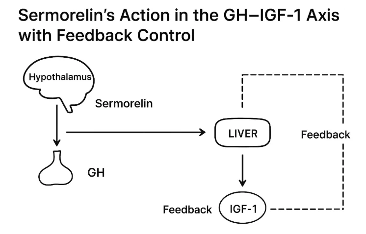
Sermorelin binds to GHRH receptors in the pituitary, activating adenylyl cyclase and increasing cyclic AMP (cAMP) levels. This triggers the synthesis and pulsatile release of Growth Hormone, which then stimulates the liver and other tissues to produce insulin-like growth factor-1 (IGF-1).¹²
Why is Sermorelin Popular in Research?
Growth Hormone Stimulation: Mimics natural GHRH to trigger pulsatile GH release in adults and children with intact pituitary function.⁴ ⁷ ⁸ ⁹
Body Composition & Aging: Restores youthful GH rhythms and improves lean-to-fat ratios in older adults without excessive IGF-1 elevation.¹⁰
GH Deficiency Research: Studied as both a diagnostic agent and potential treatment for pediatric GH deficiency; well-tolerated in clinical use.¹¹
Mechanistic Insight: Serves as a reference model for studying how the brain and pituitary regulate GH secretion over the lifespan.¹²
Summary
Sermorelin (GHRH 1-29 NH₂) is a synthetic fragment of growth hormone–releasing hormone that stimulates the pituitary to secrete GH naturally. Discovered during the early mapping of human GHRH, it became a pivotal research tool for understanding GH regulation, sleep-related secretion, and aging. Although its use as a therapy waned with the advent of recombinant GH, sermorelin remains scientifically significant — offering a window into how the brain and pituitary coordinate growth hormone release throughout life.¹-¹²
FAQs About Sermorelin
What is Sermorelin?
Sermorelin is a synthetic growth hormone–releasing hormone analog that stimulates the pituitary gland to release growth hormone in a natural, pulsatile manner.
How does Sermorelin work?
Sermorelin binds to GHRH receptors on pituitary somatotrophs, triggering cyclic AMP production and pulsatile growth hormone release.
Is Sermorelin approved for human use?
Sermorelin is not approved for human use outside of sanctioned clinical research. It is available for research purposes only.
Rivier J, Spiess J, Thorner MO, Vale W. Characterization of a growth hormone-releasing factor from a human pancreatic islet tumour.Nature. 1982;300(5889):276-278. https://pubmed.ncbi.nlm.nih.gov/6292724/
Guillemin R, Brazeau P, Bohlen P, et al. Growth hormone-releasing factor from a human pancreatic tumor that caused acromegaly.Science. 1982;218(4572):585-587. https://pubmed.ncbi.nlm.nih.gov/6812220/
Thorner MO, Rivier J, Spiess J, Vance ML, Vale W. Human pancreatic growth-hormone-releasing factor (hpGRF-40) selectively stimulates GH secretion in normal men.Lancet. 1983;1(8321):24-28. https://pubmed.ncbi.nlm.nih.gov/6129370/
Barron JL, Hopkins KD, Dunger DB, Hesp R, White A. GHRH(1-29)-NH₂ and a D-Ala² analog are potent stimulators of GH release in normal men.Clin Endocrinol (Oxf). 1985;23(4):399-407. https://pubmed.ncbi.nlm.nih.gov/2866496/
Kopelman PG, Noonan K, Ginsburg J, White N. Low-dose GHRH(1-29)-NH₂ bolus and pulsed infusions: GH responses in normal-weight vs obese women.Clin Sci (Lond). 1986;70(5):531-538. https://pubmed.ncbi.nlm.nih.gov/2871950/
Vance ML, Kaiser DL, Frohman LA, et al. [Nle²⁷]GHRH(1-29)-NH₂ in normal men: IV, SC, and intranasal dosing stimulates GH without adverse effects.J Clin Endocrinol Metab. 1986;62(6):1242-1247. https://pubmed.ncbi.nlm.nih.gov/3096623/
Losa M, Schopohl J, von Werder K. Stimulation of GH with human GRF1-44, GRF1-40, and GRF1-29 in normal subjects (single-dose comparison).Klin Wochenschr. 1984;62(23):1109-1113. https://pubmed.ncbi.nlm.nih.gov/6240568/
Merimee TJ, Furlanetto R, et al. Pulsatile GH secretion induced by GHRH(1-29)-NH₂ (sermorelin) in man.J Clin Endocrinol Metab. 1988;66(3):541-544. https://pubmed.ncbi.nlm.nih.gov/3125487/
Spoudeas HA, Hindmarsh PC, Matthews DR, Brook CGD. Low-dose GRF(1-29)-NH₂ tests in adults: dose–response characteristics.Clin Endocrinol (Oxf). 1994;40(5):583-590. https://pubmed.ncbi.nlm.nih.gov/7921207/
Khorram O, Veldhuis JD, Iranmanesh A, et al. Nightly [Nle²⁷]GHRH(1-29)-NH₂ for 4 months in older adults: activation of the somatotropic axis and body-composition effects.J Clin Endocrinol Metab. 1997;82(5):1472-1479. https://pubmed.ncbi.nlm.nih.gov/9141536/
Prakash A, Goa KL. Sermorelin: review of diagnostic and therapeutic use (pediatric GH deficiency; dosing/response).BioDrugs. 1999;12(6):419-436. https://pubmed.ncbi.nlm.nih.gov/18031173/
The Cellular Battery that Powers Energy, Repair & Longevity Research
Disclaimer: Information provided is for research and educational purposes only. NAD⁺ is not approved by the FDA or any regulatory agency for therapeutic or cosmetic use.
Introduction
Nicotinamide adenine dinucleotide (NAD⁺) is a molecule found in every living cell, essential for cellular metabolism – the process of converting nutrients into energy.³–¹² NAD⁺ acts as a cellular battery, shuttling electrons through the mitochondria to produce ATP, the body’s core energy source.¹–³ Beyond cellular metabolism, NAD⁺ also fuels enzymes that regulate cellular repair and longevity, including sirtuins and PARPs.⁴ ⁸ ⁹
Over time, these processes use up NAD faster than it can be replenished, leading to the age-related decline in NAD+ levels associated with slower metabolism, mitochondrial dysfunction, and diminished cellular resilience.⁷–⁹ Studies suggest that restoring NAD⁺ may enhance mitochondrial performance, metabolic balance, and markers of healthy aging, making it one of the most closely studied molecules in modern longevity science.¹⁰ ¹²
NAD⁺ Fast Facts
Full Name: Nicotinamide Adenine Dinucleotide
Form: Coenzyme (oxidized form NAD⁺, reduced form NADH)
Found in: All living cells (bacteria, plants, animals, humans)
Primary Role: Energy metabolism, DNA repair, cellular signaling, epigenetic regulation
Research Focus: Anti-aging, energy enhancement, cognitive support, mitochondrial function¹ ² ³
Chemical Structure
NAD⁺ is a dinucleotide coenzyme composed of two linked molecules — one containing adenine and the other nicotinamide, a form of vitamin B₃. This structure allows NAD⁺ to alternate between oxidized (NAD⁺) and reduced (NADH) states, transferring electrons to power metabolism. Because it derives from niacin, NAD⁺ directly connects vitamin B₃ nutrition to cellular energy production.
Above: NAD+ Chemical Structure. Nicotinamide (top) connected to adenine (bottom) via two ribose sugars and phosphate groups (left). Note the N in the nicotinamide structure is positively charged (N+), which tells us that it is oxidized form (NAD+) and not reduced form (NADH), in which there would N bonded to a hydrogen atom.
Discovery & Research Milestones
The story of NAD⁺ begins over a century ago. Early biochemists noticed a mysterious “co-ferment” that made yeast ferment sugars more efficiently — a clue that energy transfer depended on a small, reusable molecule. From that simple observation grew a century of research revealing NAD⁺ as one of biology’s most indispensable molecules.
Year
Study & Source
Key Finding
Early 1900s – Discovery
Harden A, Young WJ. Proc R Soc B. 1906¹
Identified an unknown “coferment” required for yeast fermentation — the first evidence of NAD⁺.
1930s – Characterization
Warburg O, Christian W. Biochem Z. 1936²
Defined NAD⁺ as a central electron carrier in cellular respiration and redox reactions.
1950s–1980s – Pathway Mapping
Preiss J, Handler P. J Biol Chem. 1958³
Traced NAD⁺ biosynthesis from niacin and described the Preiss–Handler and salvage pathways.
2000 – Enzyme Regulation
Imai S et al. Nature. 2000⁴
Linked NAD⁺ to sirtuin activation, introducing its role in gene regulation and longevity.
2004–2007 – Precursor Discovery
Bieganowski P, Brenner C. Cell. 2004⁵; Tempel W et al. PLoS Biol. 2007⁶
Identified and structurally mapped the NRK pathway, showing how nicotinamide riboside and NMN boost NAD⁺.
2010–2016 – Aging & Metabolic Decline
Houtkooper RH et al. Endocr Rev. 2010⁷; Camacho-Pereira J et al. Nat Med. 2016⁹
Demonstrated that NAD⁺ levels fall with age, contributing to mitochondrial dysfunction and metabolic disorders.
2018-2021 – Recent Research
Braidy N et al. Antioxid Redox Signal. 2018¹¹; Yoshino M et al. Science. 2021¹²
Demonstrated that NAD⁺ precursors are bioavailable and may enhance insulin sensitivity and muscle metabolism in human studies.
Summary
NAD⁺ is a universal metabolic cofactor that connects nutrition, energy metabolism, and cellular resilience. Once known only for its role in redox chemistry, it’s now recognized as a central regulator of aging and stress physiology. Ongoing studies continue to explore NAD⁺-boosting strategies to better understand how metabolism, repair, and longevity are intertwined.⁴⁵⁶⁹¹²
FAQs About NAD plus
What is NAD⁺?
“NAD⁺ (nicotinamide adenine dinucleotide) is a coenzyme found in all living cells that supports energy metabolism, DNA repair, and cellular signaling.
What does NAD⁺ do in the body?
NAD⁺ helps produce energy (ATP) in cells, activates longevity proteins (sirtuins), and supports DNA repair and mitochondrial health.
What are NAD⁺ precursors?
NR (nicotinamide riboside) and NMN (nicotinamide mononucleotide) are common NAD⁺ precursors used in research and supplements to boost NAD⁺ levels.
Why is NAD⁺ important for aging?
NAD⁺ declines with age, which may affect energy, DNA repair, and cellular health. Restoring NAD⁺ levels is a focus of longevity research.
Warburg O, Christian W. Über die Wirkung von Nicotinamid und verwandten Verbindungen auf die Gärung.Biochem Z. 1936;287:291–328.
Preiss J, Handler P. Biosynthesis of diphosphopyridine nucleotide. II. Enzymatic aspects.J Biol Chem. 1958;233(2):493–500. https://pubmed.ncbi.nlm.nih.gov/13563527/
Imai S, Armstrong CM, Kaeberlein M, Guarente L. Sir2 proteins are NAD-dependent histone deacetylases.Nature. 2000;403:795–800. https://pubmed.ncbi.nlm.nih.gov/10693811/
Bieganowski P, Brenner C. Discoveries of nicotinamide riboside as a nutrient and in NAD⁺ metabolism.Cell. 2004;117(4):495–502. https://pubmed.ncbi.nlm.nih.gov/15137942/
Houtkooper RH, Cantó C, Wanders RJ, Auwerx J. The Secret Life of NAD⁺: An old metabolite controlling new metabolic signaling pathways.Endocr Rev. 2010;31(2):194–223. https://pmc.ncbi.nlm.nih.gov/articles/PMC2852209/
Aksoy P, White TA, Thompson M, Chini EN. Regulation of intracellular NAD levels: CD38 as a major NADase.Biochem Biophys Res Commun. 2006;345(4):1386–1392. https://pubmed.ncbi.nlm.nih.gov/16730329/
Camacho-Pereira J, Tarragó MG, Chini CC, et al. CD38 dictates age-related NAD decline and mitochondrial dysfunction.Nat Med. 2016;22(10):1199–1206. https://pmc.ncbi.nlm.nih.gov/articles/PMC4911708/
Trammell SAJ, Schmidt MS, Weidemann BJ, et al. Nicotinamide riboside is uniquely and orally bioavailable in mice and humans.Nat Commun. 2016;7:12948. https://pubmed.ncbi.nlm.nih.gov/27721479/
What are the benefits and uses of Ipamorelin according to published research? As a highly selective growth hormone secretagogue (GHS), Ipamorelin is studied for its ability to stimulate growth hormone (GH) release, support body composition, enhance tissue repair, and serve as part of peptide stacking protocols (often with CJC-1295).¹²³
Disclaimer: Ipamorelin is for research and educational use only. It is not approved for human use or therapy.
Summary Table: Ipamorelin Benefits & Evidence
Application/Benefit
Evidence Level
Study Type
Notes
Growth hormone stimulation¹²
Strong preclinical, human
Animal, limited clinical
Increases GH without major effect on other hormones
Muscle & tissue repair²
Strong preclinical
Animal
Promotes recovery and regeneration
Body composition¹³
Moderate preclinical
Animal
Increases lean mass, reduces fat
Anti-catabolic effects²
Moderate preclinical
Animal
Reduces muscle wasting
Stacking (with CJC-1295)⁴
Early clinical
Human (pilot)
May enhance GH pulse and duration
Low side effect profile¹²
Clinical/preclinical
Animal, human
Minimal impact on prolactin/cortisol
Major Research-Backed Benefits
1. Growth Hormone Stimulation
Ipamorelin’s primary mechanism is the selective release of growth hormone from the pituitary gland.¹² Human and animal studies show it increases GH with minimal impact on prolactin, ACTH, or cortisol—making it attractive for researchers seeking a “clean” GH secretagogue.
2. Muscle & Tissue Repair
Preclinical data show Ipamorelin can accelerate muscle and connective tissue recovery after injury.² Why it matters: GH is crucial for cell proliferation, protein synthesis, and tissue healing
3. Body Composition
Animal research demonstrates that Ipamorelin administration can increase lean muscle mass while reducing body fat.¹³ Why it matters: Points to potential applications in studies on aging, metabolic health, or performance
4. Anti-Catabolic Effects
Ipamorelin helps blunt muscle breakdown (catabolism), making it of interest in research on muscle wasting or cachexia.²
5. Stacking With CJC-1295
Early pilot studies in humans suggest that combining Ipamorelin with CJC-1295 produces a synergistic effect on GH pulse amplitude and duration.⁴ Why it matters: “CJC-1295/ipamorelin” protocols are popular in advanced peptide research.
6. Favorable Safety Profile
Unlike older GHS peptides, Ipamorelin shows minimal risk of increasing prolactin, cortisol, or other unwanted hormones.¹²
Key benefits of Ipamorelin peptide research
Limitations & Research Gaps
Most data is preclinical; human studies are limited and small.
Long-term effects, optimal protocols, and stacking benefits require more research.
All uses are investigational—no FDA approval for any condition.
Frequently Asked Questions (FAQs)
What are the main research benefits of Ipamorelin?
Selective GH stimulation, muscle/tissue repair, body composition improvement, anti-catabolic effects, and synergistic stacking potential.
Does Ipamorelin increase other hormones like prolactin or cortisol?
No, clinical and preclinical studies show little to no effect on prolactin, ACTH, or cortisol—unlike some older GHS peptides
Why do researchers stack Ipamorelin with CJC-1295?
To amplify the pulse and duration of GH secretion, potentially enhancing overall benefits in research models.
Is Ipamorelin effective for fat loss or muscle gain?
Animal studies suggest improvements in both, but more robust human trials are needed.
Is Ipamorelin safe?
Published research reports a low side effect profile, especially compared to earlier GHS peptides.
Sermorelin is a synthetic growth hormone–releasing hormone (GHRH) analog studied for its ability to stimulate natural, pulsatile growth hormone (GH) release.¹² In research contexts, this controlled activation of the GH–IGF-1 axis has been associated with multiple physiological effects — from changes in body composition to endocrine health markers.
For research use only — not for human use. All potential benefits discussed are based on published research in animals, cells, and limited clinical studies.
Key Benefits Observed in Research
1. Stimulates Physiologic Growth Hormone Release
Sermorelin binds to GHRH receptors in the pituitary, leading to short bursts of GH secretion.¹ Unlike direct GH administration, it maintains natural negative feedback control through somatostatin and IGF-1 levels.
Why this matters: Mimicking physiologic GH rhythms may reduce the risk of side effects associated with constant hormone exposure.
2. Supports Lean Body Mass in Research Models
Animal and limited human studies show that GH pulses induced by Sermorelin can promote protein synthesis and muscle fiber repair, leading to favorable changes in body composition.³
Why this matters: Lean mass preservation is a key endpoint in aging and muscle recovery research.
3. Potential Recovery and Tissue Support
GH and IGF-1 influence connective tissue remodeling, collagen synthesis, and post-injury recovery.⁴ Sermorelin’s indirect stimulation of these pathways has been explored in models of muscle and tendon repair.
Why this matters: Recovery studies can help clarify how GH axis modulation affects healing dynamics.
4. Sleep Quality and Hormone Regulation
Some research suggests that enhanced GH pulses may correlate with improved slow-wave (deep) sleep, a phase linked to hormone balance and tissue repair.⁵Why this matters: Sleep quality is a critical variable in both endocrine research and performance recovery studies.
5. Diagnostic Tool in Endocrinology Research
Originally, Sermorelin was used in GH stimulation tests to evaluate pituitary function.² Even today, it is valuable in research protocols aimed at mapping GH responsiveness.
Why this matters: This makes Sermorelin unique among peptides — with both functional and diagnostic potential.
Limitations & Research Gaps
Most data come from animal models and short-term human studies.
Long-term safety and efficacy in specific research populations remain underexplored.
Effects may vary depending on timing, dose, and baseline endocrine function
Sermorelin acts at the pituitary, triggering GH release and IGF-1 production.
Summary
In research, Sermorelin’s benefits stem from its ability to stimulate GH naturally, supporting lean mass, tissue recovery, and hormone balance while preserving feedback control. While findings are promising, more studies are needed to confirm its long-term role in various research models.
FAQs About Sermorelin Benefits
What are the benefits of Sermorelin in research?
Sermorelin may stimulate physiologic growth hormone release, support lean mass, aid tissue recovery, and serve as a diagnostic tool in GH axis research.
Does Sermorelin improve recovery?
Research suggests that GH pulses induced by Sermorelin may support connective tissue repair and collagen synthesis, potentially aiding recovery.
Can Sermorelin be used for GH testing?
Yes. Sermorelin has been used as a GH stimulation agent to assess pituitary function in research settings.
Thorner MO, et al. Sermorelin: a growth hormone–releasing hormone analog. J Clin Endocrinol Metab. 1986;62(4):648–653. https://pubmed.ncbi.nlm.nih.gov/3004674/
Walker RF, et al. Stimulation of growth hormone secretion by Sermorelin in humans. Endocr Rev. 1994;15(1):1–14. https://pubmed.ncbi.nlm.nih.gov/8156948/
Veldhuis JD, et al. Hormonal mechanisms of muscle protein metabolism in aging. J Endocrinol Invest. 2005;28(9):S86–S92. https://pubmed.ncbi.nlm.nih.gov/16382192/
Doessing S, et al. Growth hormone stimulates tendon collagen synthesis in humans. J Appl Physiol. 2010;108(3):625–632. https://pubmed.ncbi.nlm.nih.gov/20044472/
Van Cauter E, et al. Roles of sleep and circadian rhythms in growth hormone regulation. J Clin Invest. 2000;107(2):163–168. https://pubmed.ncbi.nlm.nih.gov/10637265/
What are the benefits and uses of NAD⁺, according to current scientific research? Nicotinamide adenine dinucleotide (NAD⁺) is critical for cellular health, energy metabolism, and longevity. As NAD⁺ naturally declines with age, researchers have explored its supplementation and precursors to maintain vitality and support healthy aging.¹²³
Disclaimer: NAD⁺ supplementation discussed here is strictly for educational and research purposes. NAD⁺ products are not FDA-approved for therapeutic use.
Summary Table: NAD⁺ Benefits & Research Evidence
Benefit/Application
Evidence Level
Study Type
Notes
Energy Metabolism¹²³
Strong
Human, Animal
Critical for ATP production
Anti-aging & Longevity¹²³
Strong preclinical
Animal, Human
Sirtuin activation, mitochondrial health
DNA Repair & Cellular Protection¹²³
Strong preclinical
Animal, Cell
Enhances PARP enzyme function
Cognitive Function & Brain Health¹²
Moderate clinical
Human, Animal
Supports neuronal health
Metabolic Health¹²
Moderate clinical
Human, Animal
Supports insulin sensitivity
Cardiovascular Health¹²
Moderate preclinical
Animal
Reduces inflammation & oxidative stress
Muscle & Exercise Recovery¹²
Moderate clinical
Human, Animal
Improves muscle function & recovery
Immune Function¹²
Preliminary
Animal, Cell
May support healthy immune aging
Major Research-Backed Benefits of NAD⁺
1. Energy Metabolism
NAD⁺ is a cornerstone of cellular energy production, playing a pivotal role in converting nutrients into ATP—the cellular fuel critical for maintaining energy levels, metabolic function, and overall cellular vitality.¹²³ Why it matters: Efficient ATP production is essential for energy, performance, and healthy aging
2. Anti-aging & Longevity
NAD⁺ activates sirtuins—proteins strongly associated with healthy aging, longevity, and metabolic regulation. Animal and preliminary human studies have shown increased NAD⁺ improves mitochondrial function, reduces oxidative stress, and extends lifespan in various models.¹²³ Why it matters: Potential natural approach to longevity and healthier aging.
3. DNA Repair & Cellular Protection
NAD⁺ supports the activation of PARP enzymes, essential for repairing DNA damage and protecting genomic integrity. Higher NAD⁺ levels correlate with enhanced DNA repair, cellular resilience, and reduced cellular aging.¹²³ Why it matters: Protects cells from age-related damage and stress.
4. Cognitive Function & Brain Health
Research indicates NAD⁺ supplementation and increased NAD⁺ levels support brain health, cognition, and neuronal survival. Clinical trials and animal models suggest benefits for cognitive function, neuroprotection, and potential neurodegenerative conditions.¹² Why it matters: May support healthy brain aging and cognitive performance.
5. Metabolic Health
Clinical studies have found that NAD⁺ and its precursors (NR, NMN) improve metabolic markers, such as insulin sensitivity, weight control, and mitochondrial function—highlighting its promise for supporting metabolic health.¹² Why it matters: Crucial for managing healthy metabolism and age-related metabolic conditions
6. Cardiovascular Health
Animal research indicates NAD⁺ may improve cardiovascular function by reducing oxidative stress and inflammation, potentially offering protective effects on the heart and vascular system.¹² Why it matters: Supports cardiovascular health and longevity.
7. Muscle & Exercise Recovery
Clinical research has observed that NAD⁺ precursors enhance muscle performance, endurance, and post-exercise recovery, suggesting its role in physical performance and muscle maintenance.¹² Why it matters: May improve muscle function, recovery, and overall physical performance.
8. Immune Function
Preliminary evidence suggests NAD⁺ could support healthy immune function, potentially improving resilience and immune response during aging.¹² Why it matters: May promote healthy immune aging and resilience.
Limitations & Research Gaps
While animal and cellular data is robust, comprehensive human clinical data is still emerging.
Long-term human supplementation studies remain limited.
Precise dosage and optimal administration routes (oral vs. IV vs. injection) require further clarity.
Frequently Asked Questions (FAQs)
What are the most research-backed benefits of NAD⁺ supplementation?
Energy metabolism, anti-aging, DNA repair, cognitive support, metabolic health, and exercise recovery have strong scientific support.
Does NAD⁺ improve energy levels and metabolism?
Yes, NAD⁺ is central to cellular ATP production, directly supporting energy metabolism, mitochondrial function, and overall vitality.
Can NAD⁺ supplementation slow aging?
Research indicates NAD⁺ supports longevity pathways (sirtuins, mitochondria, DNA repair), potentially delaying aging processes. Human studies are ongoing.
What’s the difference between NAD⁺, NMN, and NR supplements?
NMN and NR are NAD⁺ precursors used in supplements because they are more bioavailable and stable. Cells convert NMN and NR efficiently into NAD⁺
Are there known risks or side effects from NAD⁺ supplementation?
Clinical studies report minimal side effects. Long-term safety at higher doses remains under research.
Verdin E. NAD⁺ in aging, metabolism, and neurodegeneration. Science. 2015;350(6265):1208–1213. https://www.science.org/doi/10.1126/science.aac4854
Yoshino J, Baur JA, Imai S. NAD⁺ intermediates: The biology and therapeutic potential of NMN and NR. Cell Metab. 2018;27(3):513–528. https://pubmed.ncbi.nlm.nih.gov/29474950/
Covarrubias AJ, Perrone R, Grozio A, Verdin E. NAD⁺ metabolism and its roles in cellular processes during ageing. Nat Rev Mol Cell Biol. 2021;22(2):119–141. https://pubmed.ncbi.nlm.nih.gov/33230262/
You must be over 21 to visit this site. By clicking “Yes”, you confirm that you are 21 years of age or older, agree that any products purchased will be used strictly for research purposes and accept our Terms of Service.